The human nose has a complex structure, its constituent elements are located both on the surface of the face and in its inner part. The nasal cavity is the initial section of the respiratory system, and the olfactory organ is also located in it. The anatomy of the organ assumes constant interaction with the external environment through the transport of air streams, therefore it is also an element of the body's defense against foreign particles and pathogenic microflora.
The structure of the nasal chamber
 The nasal cavity (cavum nasi or cavitas nasi) is the space in the middle of the upper part of the facial skull, which is located between the pear-shaped apertures and choans in the sagittal direction.
It can be conditionally divided into three segments:
- vestibule (located inside the wings of the nose);
- the respiratory area (covers the space from the bottom to the middle nasal concha);
- olfactory region (located in the upper posterior sector).
The space begins with the vestibule, which is covered with a flat epithelium and is a skin tucked inward, covering the sensory organ, retaining all its functions and having a width of 3-4 mm. On the eve there are sebaceous glands and bristle hair follicles, their intensive growth occurs. On the one hand, thanks to the hairs, large particles that come with the air are captured, on the other, the prerequisites for the development of sycosis and boils are created. The rest is covered with mucous membranes.
The septum (septum nasi) divides the nasal cavity into two unequal parts, since relatively rarely the dividing plate is located strictly in the center, more often it is rejected in one direction or another (according to various data, in 95% of the population).
Due to the presence of the baffle, the air flow is divided into equal flows.
This contributes to its linear movement and the creation of the necessary conditions for the organ to perform its main tasks (cleansing, moisturizing and warming).
In the anatomy of the septum, three areas are distinguished:
- Membranous. Small in size and most mobile, it is located between the lower edge of the cartilaginous plate and the edge of the nostrils.
- Cartilaginous. The largest in size, it has the shape of an irregular rectangular plate. The posterior upper edge joins the angle between the vomer and the ethmoid plate, the upper anterior and lateral edges - to the nasal and palatine bones, respectively.
- Bone. Formed by a number of adjacent bones (frontal, ethmoid, vomer, sphenoid, upper jaw ridges).
Newborn babies have a membrane-like septum that hardens and fully forms by about 10 years of age.
The nasal cavity, more precisely, each half of it, is limited by five walls:
- Upper (vault). It is formed by the inner surface of the nasal bones, frontal, ethmoid (with 25-30 holes for arteries, veins and olfactory nerve filaments) and sphenoid bones.
- Lower. This is a bony palate, which includes the maxillary process and the horizontal plate of the palatine bone, with incomplete or improper fusion, defects appear (cleft lip, cleft palate). Separates the nasal cavity from the oral cavity.
- Lateral. It has the most complex anatomy, it is a volumetric system of a number of bones (nasal, upper jaw, lacrimal, ethmoid, palatine and wedge-shaped), which are connected to each other in different configurations.
- Medial. This is a nasal septum that divides the common chamber into two sections.
- Back. It is present only in a small area above the choans; it is represented by a sphenoid bone with a paired opening.
The immobility of the walls of the space provides full circulation of air in it, its muscle component is poorly developed.
The nasal cavity is connected by channels with all adjacent air bones containing the paranasal sinuses (wedge-shaped, maxillary, frontal and ethmoid labyrinths).
 On the lateral wall there are nasal conchas, which look like horizontal plates located one above the other. The upper and middle are formed by the ethmoid bone, and the lower is an independent osteostructure. These shells form the corresponding paired passages under them:
- Lower. Located between the lower sink and the bottom of the chamber. In its vault, approximately 1 cm from the end of the shell, there is an opening of the nasolacrimal duct, which is formed at the birth of a child. If the opening of the canal is delayed, then the development of cystic expansion of the duct and narrowing of the passages is possible. Through the lumen of the duct, fluid flows from the voids of the eye orbit. This anatomy leads to increased mucus separation during crying and, conversely, lacrimation with a runny nose. It is most convenient to puncture the maxillary sinus through a thin section of the stroke wall.
- Average. It is located between the lower and middle shells, runs parallel to the lower one, but much wider and longer than it. The anatomy of the lateral wall is especially complex here and consists not only of bone, but also of "fountains" (fontanelles) - a kind of duplication of the mucous membrane. There is also a crescent (semilunar) gap, here through the maxillary cleft the maxillary sinus opens. In its posterior section, the semilunar slit forms a funnel-shaped expansion, through which it connects with the openings of the lattice anterior cells and the frontal sinus. It is along this path that the inflammatory process with a cold passes to the frontal sinus, and frontal sinusitis develops.
- Upper. The shortest and narrowest, located only in the posterior parts of the chamber, has a backward and downward direction. In its anterior segment it has an outlet of the sphenoid sinus, and in its posterior segment it reaches the palatine opening.
The space between the nasal septum and the turbinates is called the "common nasal passage". Under the shell of its anterior portion (about 2 cm behind the nostrils), the incisal canal emerges, containing the nerve and blood vessels.
In children, all passages are relatively narrow; the inferior shell is lowered almost to the bottom of the chamber. Because of this, almost any catarrhal inflammation and swelling of the mucous membrane lead to a narrowing of the canal, which creates problems with breastfeeding, which is impossible without nasal breathing. Also, younger children have a short and wide Eustachian tube, so when sneezing or improperly blowing their nose, infected mucus is easily thrown into the middle ear, and acute otitis media develops.

The blood supply is carried out through the branches of the external carotid artery (lower posterior region) and the internal carotid artery (upper anterior region). The outflow of blood is produced through the accompanying venous plexus associated with the ophthalmic and anterior facial veins. The specificity of blood flow often leads to intracranial and orbital rhinogenic complications. In front of the nasal septum is a small section of the superficial capillary network called the Kisselbach zone or bleeding zone.
Lymphatic vessels form two networks - deep and superficial. They both target the deep cervical and submandibular lymph nodes.
Innervation is divided into the following types:
- secretory - through the fibers of the parasympathetic and sympathetic nervous system;
- olfactory - through the olfactory epithelium, olfactory bulb and central analyzer;
- sensitive - through the trigeminal nerve (first and second branch).
Features of the structure of mucous membranes
Almost all the walls of the space, except for the vestibule, are lined with a mucous membrane, on average there are about 150 glands per 1 square centimeter of integument. The entire space can be subdivided into two sectors:
- Respiratory (lower half of the space). Covered with a cylindrical multi-row ciliated epithelium with numerous filamentous cilia that flicker, i.e. lean quickly to one side and slowly straighten. Thus, mucus, together with bound dust and harmful particles, is excreted through the vestibule and the choanae. The membrane is thicker here, since there are many alveolar-tubular glands in the subepithelial layer that secrete mucous or serous secretions. The covering of the respiratory surface is rich in cavernous plexuses (cavernous bodies) with muscular walls, which enable the caverns to contract and better warm the passing air stream.

- Olfactory (upper shells and half of the middle shells). Its walls are covered with pseudo-stratified epithelium, which contains bipolar neurosensory cells that sense odors. Their front side bubbles outward, where it interacts with molecules of odorous substances, and the back passes into nerve fibers, which, intertwining into nerves, transmit a signal to the brain, which recognizes aromas. In addition to the specific olfactory layer of the epithelium, there are cylindrical cells, however, devoid of cilia. The glands in this area secrete a liquid secretion for hydration.
In general, the lamina of the mucous membrane, despite some differences, is thin and contains, in addition to mucous and serous glands, numerous elastic fibers.
In the submucosa, there are lymphoid tissues, glands, vascular and nerve plexuses, as well as mast cells.
Functions of the nasal cavity
The nasal chamber, due to its location and anatomy, is adapted to perform a large number of the most important functions of the human body:
- Respiratory. Inhaled air travels along an arched path to the nasopharynx and back, while it is humidified, warmed and cleaned. Thin-walled veins and a large number of small blood vessels increase the air temperature. Moisturizing occurs due to the intense release of moisture by secretory cells. Also, air that is inhaled through the nose, putting pressure on the walls of the chamber, stimulates the respiratory reflex, which leads to an expansion of the chest more than with oral breathing.
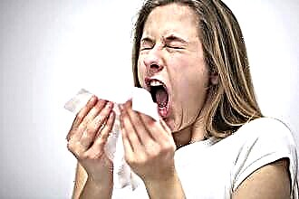 Protective. The mucus secreted by goblet cells and alveolar glands contains lysozyme and mucin, therefore it has bactericidal qualities. It has the ability to capture and bind suspended particles in the incoming air stream, viruses and pathogenic bacteria, which are subsequently excreted with the help of cilia of the ciliated epithelium into the nasopharynx through the choanae. Protection from coarse suspended particles or other airborne irritants is provided through the sneezing mechanism. This is a sharp reflex exhalation through the nostrils due to irritation of the endings of the trigeminal nerve. Also, the body is protected from harmful impurities with the help of increased secretion of the lacrimal gland, while tears are directed not only to the outer part of the eyeball, but also to the nasal chamber through the nasolacrimal duct.
- Olfactory. Recognition of odors perceived by the olfactory epithelium and sent along nerve endings to the brain for information processing.
- Resonator. Together with the sinuses, mouth and throat, they create sound resonance, giving the voice a unique individual timbre and sonority. With a runny nose, this function is partially violated, which makes the voice deaf and nasal.
 Protective. The mucus secreted by goblet cells and alveolar glands contains lysozyme and mucin, therefore it has bactericidal qualities. It has the ability to capture and bind suspended particles in the incoming air stream, viruses and pathogenic bacteria, which are subsequently excreted with the help of cilia of the ciliated epithelium into the nasopharynx through the choanae. Protection from coarse suspended particles or other airborne irritants is provided through the sneezing mechanism. This is a sharp reflex exhalation through the nostrils due to irritation of the endings of the trigeminal nerve. Also, the body is protected from harmful impurities with the help of increased secretion of the lacrimal gland, while tears are directed not only to the outer part of the eyeball, but also to the nasal chamber through the nasolacrimal duct.
Typical diseases of the nasal cavity
Diseases of the constituent parts of the space under consideration depend on many factors: structural features of each individual, disorders of certain functions of organs, exposure to pathogens or medications.
The most common ailment is a runny nose of various types:
- Acute rhinitis is an inflammation of the mucous membrane, leading to dysfunctions of the olfactory organ. It can be an independent disease or a symptom of a more general illness (flu, colds, SARS). Signs of acute rhinitis are congestion, abundant secretion, loss of smell, difficulty breathing.
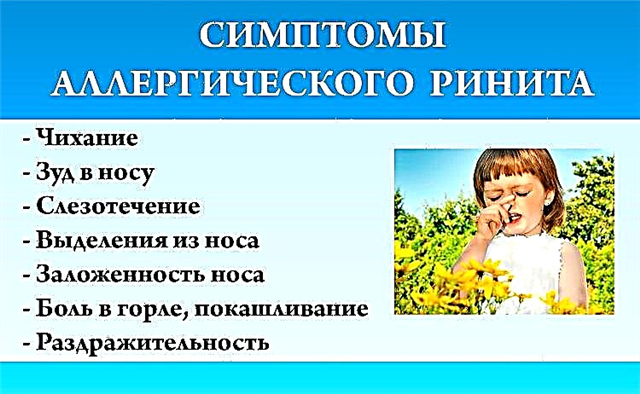
 Vasomotor rhinitis (neurovegetative or allergic) is a violation of the tone of the blood vessels of the shells due to infections, stress, hormonal disorders or an individual reaction to certain stimuli (pollen, dust, fluff, animal hair, perfume). May be permanent or seasonal. At the same time, ventilation of the lungs worsens, the patient gets tired quickly, appetite and sleep are disturbed, and headaches appear.
- Hypertrophic rhinitis. It is often a consequence of other types of rhinitis, is mainly chronic in nature and consists in the proliferation and thickening of connective tissues. Breathing in this case is constantly difficult, therefore, most often doctors recommend an operation, excising the overgrown tissue surgically.
- Atrophic rhinitis. Dystrophic changes in the epithelial membrane of the organ. It is characterized by dryness in the passages, the appearance of dried crusts, loss of smell, and breathing problems.
- Rhinitis medication occurs as a result of improper use of drugs (drops or sprays) for a long time.
 Vasomotor rhinitis (neurovegetative or allergic) is a violation of the tone of the blood vessels of the shells due to infections, stress, hormonal disorders or an individual reaction to certain stimuli (pollen, dust, fluff, animal hair, perfume). May be permanent or seasonal. At the same time, ventilation of the lungs worsens, the patient gets tired quickly, appetite and sleep are disturbed, and headaches appear.
Almost all types of rhinitis, except for hypertrophic, are amenable to conservative local treatment: irrigation, rinsing with medicinal solutions, turunda with ointments.
Other organ diseases include:
- Synechia. This is the formation of tissue adhesions, most often due to surgery or various injuries. When the problem is eliminated with a laser, relapses are rarely recorded.
- Atresia. Fusion of tissues of natural channels and openings. Most often it is congenital, but it can also be acquired, as a complication of syphilis, diphtheria. In older patients, thermal and chemical burns, abscess of the nasal septum, trauma, and unsuccessful operations also became the reasons. As a result, accreted tissues partially or completely block the nasal passage, and a person can breathe only through the mouth. After fluoroscopy, it is possible to perform an operation to form lumens.
- Ozena. Disorders of tissue nutrition due to dysfunction of nerve endings, degeneration of the epithelium, which disintegrates and emits a fetid odor that is not felt by the patient due to the death of the olfactory receptor. The nose is very dry, and the crusts can clog the passages, although they are very dilated. The disease is still not well understood.
- Polyps. Chronic rhinosinusitis, changing the structure of the epithelium, can lead to the development of polyposis. It is usually treated promptly by destroying the leg of the polyp.
- Neoplasms. These may include papillomas, osteomas, cysts, fibromas. The strategy of their treatment is developed for each specific case, taking into account the data of additional studies.

- Injuries. Most often, there is a curvature of the nasal septum due to bone fracture or improper fusion. In addition to a cosmetic problem, in such cases, nighttime snoring, drying out, bleeding is observed, sinusitis, frontal sinusitis, allergic reactions can develop, immunity deteriorates and susceptibility to infections increases. The defect is corrected surgically.
Doctors recommend starting treatment of any nasal diseases immediately, since the resulting lack of oxygen negatively affects all body systems, oxygen starvation is especially dangerous for the brain. Switching to mouth breathing does not solve the problem, but only exacerbates it. Shortness of breath through the mouth:
- The entry into the lungs of unmoistened and unheated air.Less efficient gas exchange occurs in the alveoli, less oxygen enters the bloodstream.
- The body's defenses are weakened due to the exclusion of mucus from the process, the risk of respiratory infections increases dramatically.
- Long-term mouth breathing contributes to inflammation of the pharyngeal tonsil - adenoiditis.
Techniques for examining the nasal chambers
In order to identify the disease and determine the stage of its development, the following basic diagnostic methods are used in modern medicine:
- Anterior rhinoscopy is performed in each case using a special nasal dilator, the tip of the nose is lifted and the instrument is inserted into the nostril. Each nostril is visually inspected separately, sometimes a bulbous probe is used. On examination, problems such as inflammation of the walls, curvature of the septum, hematomas, polyps, abscesses and neoplasms can be detected. In case of tissue edema, the doctor first drips vasoconstrictors into the passages (for example, a 0.1% adrenaline solution). An autonomous light source or a head-mounted reflector is used to illuminate the surveyed area.
- Posterior rhinoscopy is used when indicated. In this case, the nasopharynx and nasal cavity are examined from the side of the choanas. The doctor in the open throat with a spatula pushes the root of the tongue and inserts a special mirror with a long handle into the throat.
Additional, more specialized studies include:
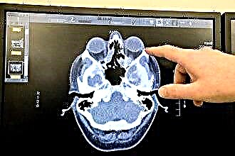 X-ray of the skull. In this case, the state of all cavities of the skull, anomalies and deformations of the bones are studied. An X-ray is taken in different projections if necessary to obtain a more voluminous picture.
- Computed tomography gives a better and more complete image than radiography. As a result of its implementation, defects of the posterior part of the nasal septum are revealed, which cannot be seen during rhinoscopy (spines and ridges).
- Endoscopy is performed using a thin probe (rhinoscope) with a microcamera at the end. After local anesthesia with anesthetic sprays, the probe is inserted through the nostril and advanced inward. Helps to identify various formations that are inaccessible with posterior and anterior rhinoscopy. Usually well tolerated by patients.
 X-ray of the skull. In this case, the state of all cavities of the skull, anomalies and deformations of the bones are studied. An X-ray is taken in different projections if necessary to obtain a more voluminous picture.
Laboratory diagnostic methods:
- A general blood test is a routine general clinical study, carried out if any disease is suspected. Allows you to determine the signs of the inflammatory process.
- Bacteriological examination of separated mucus and smears. It makes it possible to accurately determine the causative agent of the disease and choose a rational antibiotic therapy.
- Cytological examination of secretions and smears. It is used when there is a suspicion of the presence of an oncological process.
- Immunological studies and allergy tests. Identification of allergens that provoke the development of ailments.