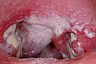
 Diseases of the human auricle are quite diverse and can be the result of inflammatory processes, infections, congenital pathologies, fungi. They are dangerous because of the location of the auditory organs and the possibility, in case of complications, to affect the brain and central nervous system. The most common diseases of the auricle in humans should be considered in more detail.
Erysipelas
Erysipelas of the auricle is an infectious disease widespread in the world, which is characterized by exudative-serous inflammation of the skin, less often of the mucous membranes. The causative agent is group A streptococcus.
Erysipelas is often preceded by streptococcal infections in acute (upper respiratory tract catarrh, tonsillitis) or chronic (periodontitis, caries, purulent sinusitis) form. You can get infected from a sick person through contact through mucous membranes or damaged skin, as well as by airborne droplets.
Symptoms of erysipelas, for which the disease is diagnosed:
- severe pain on palpation;
- swelling of the entire outer ear, including the lobe;
- a sharp increase in body temperature (up to 40 degrees);
- chills;
- burning;
- the appearance of bubbles filled with serous fluid (bullous form).
For cure, compulsory antibiotic therapy is carried out for 8-10 days with the help of drugs such as amoxicillin, cefadroxil, cefuroxime. If the patient does not tolerate beta-lactates, then alternative antibiotics are prescribed - erythromycin, spiramycin, azithromycin.
Local treatment consists in applying a 2% mupirocin ointment to the affected area, lubrication with anti-inflammatory or indifferent ointments, irradiation with an erythemal dose of ultraviolet rays. With adequate therapy, in mild cases, recovery occurs in 3-4 days.
In severe cases, it can be delayed and accompanied by exacerbation and remission processes.
Perichondritis
Perichondritis is an inflammation of the auricle, the treatment of which must be carried out in order to prevent the cartilage from melting. It begins with an infection in the perichondrium, the most common pathogens are:
- Pseudomonas aeruginosa;
- Staphylococcus aureus;
- green streptococcus.
As the disease develops, it covers the skin and the membranous part of the external auditory canal. At the initial stage, the disease is serous, eventually turning into purulent.
Bacteria enter the body through trauma to the organ of hearing, scratching, insect bites, abrasions, frostbite and burns.
At risk are people with weak immunity, taking corticosteroid drugs, diabetes mellitus.
The most characteristic signs of perichondritis:
- discomfort and pain in the ear canal;
- redness and swelling of the ears;
- burning;
- manifestation of a focus of suppuration;
- temperature rise to 38-39 degrees;
- weakness;
- loss of appetite;
- increased pain on palpation.
During the examination, a specialist must differentiate perichondritis from erysipelas and festering hematoma.
Conservative treatment is effective only for the serous form of the disease: antibiotics, sulfonamides, macrolides (josamycin, cleithromycin), physiotherapy (laser therapy, microwave, ultraviolet radiation). With purulent perichondritis, empyema is opened, pus is removed, the wound is washed with antibiotic solutions, drained and bandaged.
Chondrodermatitis nodosa
Chondrodermatitis nodosa of the auricle is a cartilage disorder in which an extremely painful papule forms at the edge of the antihelix or curl. The disease is typical for people over 40 years old, with age, the frequency of occurrence increases. In men, the area of the curl is more often affected, in women, the antihelix. The exact cause is unclear, possibly due to repeated trauma.
The initial lesion is a red painful hard papule 3-4 mm in diameter.
 In the center, there is a keratinization point covered with a crust. The surrounding skin shows signs of atrophy and actinic lesions. More often the focus is one, less often - several, very rarely - on both sides. The main symptom is a sharp stabbing pain and tenderness on palpation.
Laboratory diagnostics:
- A biopsy reveals an inflammatory process (both acute and chronic), the signs of which are a thin epidermis, erosion, and parakeratosis.
- Skin necrosis with granulation tissue.
- Cartilage degeneration with deep biopsy.
- In many ways, chondrodermatitis nodosa resembles squamous cell or basal cell carcinoma.
The therapy of the disease is rather complicated; local treatment is rarely effective. It consists in reducing pressure on the affected area (especially during sleep) and injecting steroids. To heal, it is necessary to remove the inflamed part of the cartilage along with the lesion. However, after any treatment, the relapse rate is high.
Hypertrichosis
Hypertrichosis is excessive hair growth in various parts of the body, especially those where hair growth is not predetermined by hormones. It is to such cases that hypertrichosis of the auricle belongs. The disease can affect men, women and children.
Causes of the disease:
- Congenital pathology, when epithelial cells are transformed into cells containing hair follicles. Mutations occur due to unfavorable pregnancy or infectious diseases in the first trimester of pregnancy. The mutated gene can be inherited.
- An acquired trait under the influence of various factors. For example, it can be the activity of tumor markers or climacteric changes in women.
- Medicinal. Sometimes it manifests itself after prolonged use of certain antimicrobial drugs, such as penicillin, streptomycin, corticosteroids.
- Also, hypertrichosis can provoke fungal lesions, traumatic brain injury, anorexia, scars and burns.
With an endocrine cause of the disease, therapy consists in changing the drugs taken. If the hypertrichosis is congenital, then cosmetological and aesthetic treatment is used by methods of photo- and electrolysis, these are expensive and time-consuming procedures. For children, hair is lightened with hydrogen peroxide and removed with special creams.
Deformation
Deformation of the auricles is quite common in people, the causes of which can be congenital and acquired. Damage can significantly reduce the functionality of the organ. They are often perceived as a cosmetic problem and then develop into hearing loss or otitis media.
 The causes of congenital deformity can be transferred intrauterine infections and injuries, genetic inheritance, facial anomalies. Acquired deformities are usually associated with active sports (boxing, wrestling) or domestic injuries.
There are no pronounced symptoms, most often patients complain of a deterioration in sound patency.
An otolaryngologist or traumatologist is able to determine the damage, if necessary, computed tomography is used.
The treatment is carried out in a comprehensive manner and consists in the alignment of the cartilaginous tissues and the release of the ear canal. Infections are removed by medication, after which surgical correction (ear otoplasty) is performed.
Otohematoma
Auricle hematoma in humans is mainly the result of bruising, trauma or blow. It looks like a cavity filled with liquid or coagulated blood, located between cartilage and skin or between cartilage and perichondrium. It appears as a result of trauma to an artery or vein in the ear.
The symptoms are:
- swelling and redness of the affected area;
- soreness when pressed with fingers;
- an increase in local or general temperature;
- accumulation of blood in the cavity under the skin of the organ of hearing.
The hematoma can rapidly increase in size within 2-3 days with increased pain. Then the redness and pain disappear, and the hematoma transforms into a thickening of fibrin and connective tissue. To clarify the diagnosis, the doctor can make a puncture and take part of the contents for analysis. If this cannot be done, then we can talk about an abscess.
Small hemorrhages are able to resolve on their own; it is enough to apply a tight bandage and cold. If you feel uncomfortable, take an anesthetic or anti-inflammatory drug. In more difficult cases, for example, when a hematoma forms on the front of the ear, a puncture is performed under local anesthesia, the collected blood is sucked off, the cavity is washed and drained, and antimicrobial drugs are prescribed. Without timely treatment, a large hematoma can fester and develop into perichondritis.


