Bumps on the ears of a person can have a different origin and be both an independent disease and one of the signals of more global malfunctions in the body. Inflammatory processes of a general and local nature, benign and malignant neoplasms can visually look like a lump on the ear. What it is, only an otolaryngologist is able to say for sure after a visual examination and study of the test results.
Inflammatory diseases
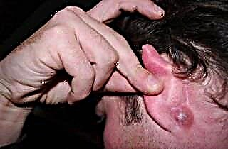
 If a lump has jumped on the ear and it hurts, has a bright red color, then this may be a sign of the formation of a boil. Boil is a purulent inflammation of the hair follicle (less often the sebaceous gland), in which a necrotic shaft is formed, surrounded by pus. The causative agent is Staphylococcus aureus, which is activated when a favorable environment is created for it.
The reasons for the development of the boil of the auditory organ can be:
- non-compliance with hygiene requirements;
- injury to the skin or hair follicle;
- frequent purulent otitis media;
- hormonal surges;
- metabolic disease;
- viral infections, colds and hypothermia;
- low immunity.
Symptoms of the disease are fever, swelling of nearby tissues, severe pain from the pressure of pus on the nerve endings, which shoots into the temple, neck, eyes, teeth. The patient does not sleep well, it is difficult for him to chew and speak. The boil develops for 3-4 days. A lump on the ear cartilage is especially painful.
 You can treat a boil with the help of means that accelerate its ripening. The most famous drugs are Vishnevsky's ointment and zinc ointment, which are applied to a tampon, applied to the affected area and fixed with a plaster or bandage. Saline solution, applied as a compress at night, also helps.
Adepts of traditional medicine recommend applying peeled aloe leaves to the boil or smearing it with Zvezdochka ointment.
To improve well-being during the development of the boil, the doctor prescribes antipyretic drugs, analgesics or antibiotics. With a sharp increase in body temperature and severe pain, the doctor recommends surgical removal of the abscess. During the operation, an abscess is opened, the necrotic rod and the surrounding exudate are removed, after which a drain is placed in the wound. The patient's condition improves dramatically the next day.
An attempt to independently open the abscess often ends with the insemination of adjacent hair follicles with pus and the development of new foci. They tend to unite into one large focus with several rods and create a carbuncle, which heals for a very long and difficult time, and after which visible scars remain.

Sometimes pineal edema can occur with the serous form of perichondritis of the auricle, a disease that affects the perichondrium. In this case, the shell becomes edematous, shiny and red. The resulting swelling gradually concentrates in a painful compaction to the touch. Perichondritis is a dangerous disease, its untimely treatment can lead to cartilage melting.
Benign tumors
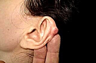
The most common hearing growths are atheroma and lipoma, which may look like a lump on the ear or lobe. In a normal state, these diseases do not cause any particular inconvenience, except for cosmetic ones, and are able to completely dissolve on their own after some time.
 Atheroma (cyst) is a clogged sebaceous gland that is severely distended due to accumulated fat. To the touch, it looks like a hard ball, which, as a rule, does not bother. However, in the case of suppuration, it can sharply increase in size and hurt, and the body temperature rises. In this case, the atheroma must be removed. At an early stage, before inflammation, it is removed with high-frequency radio waves or a laser, at a later stage - with a traditional scalpel. It is impossible to try to squeeze out on your own, since the entry of the contents of the capsule into the bloodstream can cause a sharp deterioration in the patient's condition and even sepsis.
- Lipoma (wen) is most often formed in the layer of subcutaneous tissue and is an overgrowth of fat cells. It is a soft, painless formation with clear boundaries that can move under the skin. It does not pose a health hazard, since it does not grow into neighboring tissues and does not degenerate into more dangerous forms. If necessary, it is easily removed surgically using a laser, which excises the fatty clot and coagulates the blood vessels.
 Atheroma (cyst) is a clogged sebaceous gland that is severely distended due to accumulated fat. To the touch, it looks like a hard ball, which, as a rule, does not bother. However, in the case of suppuration, it can sharply increase in size and hurt, and the body temperature rises. In this case, the atheroma must be removed. At an early stage, before inflammation, it is removed with high-frequency radio waves or a laser, at a later stage - with a traditional scalpel. It is impossible to try to squeeze out on your own, since the entry of the contents of the capsule into the bloodstream can cause a sharp deterioration in the patient's condition and even sepsis.
Also, a seal on the outside of the auditory organ can be:
- Papilloma. It is acquired and congenital, often appears on the auricle and is small in size;
 The fibroma grows from the cells of the connective tissue and is often localized at the site of the piercing;
- Nevus is a birthmark formed by pigment cells in combination with the nerve sheaths. Has a different shape, size and color;
- Hemangioma occurs when blood vessels grow together, grows deep into the shell and ear canal, destroys nearby tissues, and can cause severe bleeding;
- Chondroma occurs in the cartilaginous part of the ear and is rare;
- Osteoma. The place of its localization is the posterior wall of the bone section, it can penetrate into the mastoid process, it is dangerous with relapses.
 The fibroma grows from the cells of the connective tissue and is often localized at the site of the piercing;
Malignant tumors
Secondary tumors - a consequence of the development of metastases from oncological diseases of other organs - are treated for a long time and are difficult. Primary tumors on the ears are an independent pathology, with timely detection and initiation of therapy, the result is generally positive. The main types of malignant neoplasms:
 Melanoma. It is formed from pigment cells and quickly grows in the skin. Able to spread metastases through the lymphatic and blood channels.
- Basalioma. It develops in the upper layer of the skin, looks like a plaque or a nodule of a reddish-gray hue, and may peel off. Malignancy is low, does not metastasize.
- Spinocellular epithelioma. A widespread tumor, which is an ulcer that grows in depth and in breadth on the lobe or at the ear canal. May gradually invade the cranial cavity, salivary glands and middle ear. It is most common in older men.
- Adenocarcinoma. It is formed from glandular cells near the ear canal, can grow into the temple and the middle of the organ of hearing.
- Sarcoma. It is rare, more often in children. Located on the sink or near the ear canal.
 Melanoma. It is formed from pigment cells and quickly grows in the skin. Able to spread metastases through the lymphatic and blood channels.
Treatment of malignant neoplasms is carried out in a complex, using radiation, chemotherapy and surgical methods.
Diagnostics
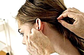
 If a lump occurs on any part of the ear, you should consult an otolaryngologist (ENT). He will conduct an external examination (otoscopy) and, if necessary, involve a dermatologist or dermato-oncologist in the diagnosis.
Further, in accordance with the indications, such in-depth diagnostic methods can be used:
- computed tomography (CT);
- radiography;
- magnetic resonance therapy (MRI);
- biopsy followed by histological analysis of the affected tissues;
- pharyngoscopy;
- audiometry.