A cyst in the maxillary sinus is diagnosed in approximately 10% of the world's population. It can be localized on the left and on the right side of the organ, most often it chooses for itself the inner lower part of the mucous membrane. The disease is not oncogenic, it does not metastasize and does not spread to other organs, therefore it is not considered very dangerous to health. However, when creating ideal conditions for its progression, very unpleasant and serious complications can appear.
What is a neoplasm
 The maxillary sinus is an accessory air cavity in the skull, it connects to the nose and actively interacts with it. The inner surface of the pocket consists of a mucous membrane that produces mucus through its glands. The secret in favorable conditions is excreted through special ducts, it performs two functions at once: moisturizes and kills pathogens. However, it is not always possible to resist viruses, bacteria and fungi, especially when there is a strong inflammatory process in the nasal cavity.
If there is constant swelling in the mucous membrane, it begins to malfunction. The ducts through which mucus is transported gradually become clogged or overgrown, and their obstruction occurs. Since all the pathways are blocked, the secret has nowhere to go, because it begins to accumulate in the glands, which gradually increase in size from the contents. Small elastic balls are formed on the mucous membrane, this is the cyst of the maxillary sinus.
Types of neoplasms
There are true and false neoplasms. The division into groups depends on the mechanism of formation of cysts and their structure. The location of the liquid bubbles is also important. During the examination, it is important to find out the type of disease in order to choose the most appropriate treatment or removal technique. Let's consider them in more detail:
- Retention cyst of the maxillary sinus (true). We discussed the mechanism of the appearance of this type of cyst above, they are formed on the mucous membrane of the maxillary pocket on the left or on the right, if the ducts, through which the secret leaves, become clogged or grow together. The peculiarity of these neoplasms is that they are two-layer, the inner part consists of epithelial tissue, which also produces mucus.
- Odontogenic cyst of the maxillary sinus (pseudocyst). Pseudocysts or false cysts have their own specific origin. They appear in the air pockets due to diseases of the teeth and gums. The main criterion for their development is infections that get from the tooth into the root canal. When the bone begins to collapse, a ball filled with liquid forms, which separates pathologically dangerous tissues from healthy ones - this is a kind of protection against the spread of infection. As the disease spreads, the cyst grows in size, it can completely destroy the bone, leaving voids in it. These neoplasms have a single layer, they can disappear on their own if the patient cures dental diseases.
Major risk factors
Neoplasms in the maxillary sinuses do not form just like that; certain conditions must be created for their appearance. Frequent irritation of the nasal mucosa and maxillary pocket is the most common cause of the disease. Patients with chronic sinusitis are especially susceptible to it. Also, not completely cured pathologies of the teeth and gums can serve as a trigger.

The main factors causing the violation:
- chronic or often recurrent inflammation of the nasal passages and paranasal sinuses;
- inflammation affecting the upper jaw and its teeth;
- constant contact with allergens with personal intolerance;
- abnormal structure of the sinus;
- drop in general and local immunity.
How does the disease manifest itself
 If a cyst in the maxillary sinus has arisen recently and its size is less than 1 cm, it may not make itself felt at all. Most patients live with this disease, and do not suspect that they have it. Small growths do not cause difficulty breathing air, pain, or other symptoms. However, if bubbles grow, they can give certain signals about their existence:
- localized pain (a cyst of the right maxillary sinus causes pain on the right side, and a cyst of the left maxillary sinus - on the left);
- pain with irradiation (radiates to the temple and orbit, occurs on the side of the affected sinus);
- pain when changing atmospheric pressure (occurs when diving to depth or flying in the air);
- Occasional or persistent nasal congestion on one side (left maxillary sinus cyst causes left nostril congestion, and right maxillary sinus cyst causes right nostril congestion);
- nasal discharge (found on the side of the lesion, can be transparent or purulent with an unpleasant characteristic odor).
To cure or not to cure?
 To date, doctors cannot come to a unanimous decision as to whether the symptom-free maxillary sinus cyst requires removal or not. The category of doctors who advocate surgical intervention is confident that with the slightest disturbances in the work of the nasal mucosa or accessory pockets, the growth of the neoplasm will increase in volume. If progression is not detected in time, complications such as depletion of the mucous membrane, spread of infection to nearby organs, and even damage to the nasal septum are possible. To prevent such consequences, doctors recommend immediately getting rid of the cyst and not waiting for its growth.
The second part of the specialists who oppose surgical measures are sure that unnecessary interference with the mucous membrane does not have a beneficial effect on the patient's health. This can lead to complications such as partial or complete loss of smell.
The maxillary cyst behaves unpredictably, it is impossible to accurately predict its growth. In some cases, it does not change its size and even disappears, but sometimes it progresses rather quickly. Those who have found asymptomatic neoplasms are shown to be examined every six months.
Subtleties of diagnosis
If a cyst has formed in the maxillary sinus, this does not mean that you will see it immediately or feel it. Most often, the disease is diagnosed completely by accident, since it has symptoms similar to many other disorders. A very common story when a patient comes with suspicion of sinusitis, and after an X-ray examination it turns out that he has a cystic neoplasm. There are a number of procedures that help determine the presence and characteristics of cysts, let's get acquainted with them.
 X-ray examination. Only large neoplasms are visible on the x-ray; they most often fill most of the paranasal sinus.
- MRI and CT. Magnetic resonance imaging and computed tomography makes it possible to detect even small neoplasms, and during research, you can find out the size of the cysts, their location, find out if there are any concomitant diseases.
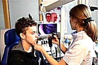
- Endoscopy. It is necessary to clarify the anatomical and physiological features of the structure of the sinuses and nasal cavity, assess the cyst itself, its size and location.
- Orthopantomogram. This is a panoramic image of the jaw, which is taken when a monotonic cyst is suspected.It allows you to see all neoplasms in the jaw, even if they have not yet relocated to the area of the maxillary sinuses.
- Biopsy. Laboratory research of a miniature piece of material (cyst) makes it possible to determine its features, to find out the type of neoplasm and even the reasons that led to its appearance.
How to get rid of
It is possible to get rid of a cyst of the maxillary sinuses, regardless of the reasons for its appearance, only with the help of an operation. Conservative treatment is sometimes used, but it only helps to relieve severe symptoms for a while. Puncture of the neoplasm is especially popular. After the liquid is removed, the bag itself does not go anywhere; over time, it refills again. Warming up and similar physiotherapy procedures are completely contraindicated, as they can aggravate the situation.
The only way to guarantee a complete recovery is surgery.
Operations can be performed using the classical method (Caldwell-Luke and Denker operation). In the first variant, trepanation of the maxillary sinus is performed through the upper gum, and in the second, through the front wall. Both techniques are quite traumatic and are rarely used in our time.
The least painful and most effective is endoscopic removal - this is the "gold standard", as the surgeons themselves say. With the help of an endoscope, you can work only with affected tissues, without affecting healthy ones. The operation is performed under local anesthesia and does not require a long period of rehabilitation and trepanning of the face.
Preventive measures
 People who often experience rhinitis and sinusitis, as well as allergy sufferers or regular patients of dentists have a predisposition to the formation of cysts of the upper sinuses. A scrupulous attitude to your health will help to avoid the appearance of cystic neoplasms.
- If you have allergies, take antihistamines on time so as not to irritate the mucous membranes.
- In the presence of acute and chronic ENT diseases, they must be treated to the end, do not ignore the drugs prescribed by the doctor.
- Any problems with the teeth, especially in the upper jaw, need to be addressed as quickly as possible, as inflammation can lead to the formation of cysts.
- Strengthening local and general immunity will also be an effective preventive measure.
In conclusion
The maxillary sinuses are a favorite location for cysts. The disease is not considered difficult or very dangerous, there is even disagreement between doctors about the removal of asymptomatic neoplasms. However, a patient who has already found this disorder must carefully monitor his health and undergo examinations in time so that the doctor can control the condition of the cyst.

 X-ray examination. Only large neoplasms are visible on the x-ray; they most often fill most of the paranasal sinus.

