Aortic valve device, its function
The aortic valve (AK) separates the left ventricle from the aorta, preventing blood from flowing back to diastole (myocardial relaxation phase). Its other name - lunar - reflects the structural features, since the AK is formed by three convex pockets (flaps).
Anatomy
AK is located in the initial section of the vessel - the aortic bulb, which is projected onto the chest in the center of the sternum, between the cartilage of the third ribs.
The structure of the AK is very complex, the valve consists of:
- three lunar flaps (or flaps);
- fibrous ring;
- commissar.
Sometimes it is supplemented with Valsalva sinuses and Henle triangles. These elements do not anatomically belong to the aorta, but take part in the work of the structure.
The annulus fibrosus consists of connective tissue bundles formed by collagen and elastic fibers. The element forms the border between the ventricle and the aortic bulb, being the point of attachment of the leaflets.
Dampers are the main functional part of the AK. In their shape, they resemble pockets that extend from the walls of the aorta, with their base attached to the fibrous ring. The free edge of each valve is slightly elongated and contains an Arrancius knot at the end.
There are three flaps: right, left and rear. Opposite each are the so-called sinuses (or sinuses) of Valsalva - the entrance gate of the coronary vessels that feed the myocardium of the heart.
Commissures - the lines of contact of the edges of the dampers at the moment when the valves are closed. The normal activity of the heart largely depends on the density of their articulation.
Histology
All valves, including the aortic valve, are formed from the endocardium - the inner lining of the heart, mainly consisting of epithelial cells. However, each structure has its own characteristics:
- The annulus fibrosus is formed by connective tissue, which gives a certain rigidity and density. The need for such a structure is caused by the high hemodynamic load that the element is subject to.
- The flaps are formed by three layers of connective tissue: fibrous (or aortic), spongy and ventricular. They contain a large amount of collagen and a small amount of elastin. Outside, each leaflet is covered with a thin endothelial membrane.
- Valsalva sinuses have a thinner wall in comparison with the aorta. The latter consists of two layers: intima and media. Towards the heart, collagen decreases and elastin increases.
During embryogenesis, AK develops from the mesenchyme, like all tissues of the left ventricle.
Physiology
The physiological importance of AK is enormous, since the valve regulates the normal flow of blood from the ventricle to the system of the great circle, which feeds the entire body. In addition, adequate valve closure is involved in filling the coronary arteries.
The valve works passively, under the influence of blood coming from the heart. The whole process is divided into two stages - the periods of opening and closing the sashes.
The opening phase contains several stages:
- Preparatory. At this moment, the heart is in a phase of isovolumetric (constant size and volume of the chamber) contraction. In this case, all the valves are closed, and during muscle tension in the left ventricle, pressure rises rapidly. In addition, the aortic root expands, with the result that the flaps begin to open even before there is a difference in pressure on both sides.
- Quick opening begins at the moment when the pressure in the ventricle exceeds the value in the aorta, after which blood rushes out of the heart, pressing through the flaps.
- Opening peak coincides with the rapid expulsion phase. The flaps at this time are tightly pressed against the sinuses of Valsalva, the lumen approaches the shape of a circle.
The closing period consists of two phases:
- Sustainable discovery corresponds to the slow ejection phase. The pressure begins to equalize, the flaps partially move away from the walls, the lumen looks more like a triangle shape.
- Fast closing. Due to the slow blood flow, small turbulences are formed near the walls. Reaching the sinuses, they penetrate the valve pockets and move the leaflets towards the center, thereby closing them.
Swinging shut, the flaps emit a sound, which, upon auscultation, is recorded as the 2nd tone. Additional noise comes from reverse blood flow during diastole when fluid strikes a closed valve.
Basic valve pathologies and methods of their correction
There are congenital and acquired diseases of AK. The first category includes the following pathologies:
- A bivalve valve is a dangerous condition in which sclerosis and adhesions develop between the flaps. Pathology leads to stenosis (narrowing of the lumen) and progressive AK dysfunction, which requires surgical intervention.
- The four-leaf valve is characterized by incomplete closure of the leaflets, which leads to insufficiency and reverse flow of blood.
Congenital malformations are caused by the interaction of genetic mutations (for example, Marfan syndrome) and external factors in the prenatal period (toxic substances, medications, radiation or maternal illness).
Acquired anomalies develop under the influence of:
- autoimmune diseases (rheumatism, systemic lupus erythematosus, Paget's disease);
- atherosclerosis;
- metabolic cardiomyopathies (toxic, with diabetes mellitus or thyroiditis);
- infectious pathologies (syphilis, bacterial myocarditis).
Long-term damage results in stenosis or insufficiency (associated with regurgitation) of the valves.
AK stenosis is a narrowing of the lumen between the flaps due to their fusion, as a result of which the outflow of blood from the heart to the aorta becomes difficult. In this case, hypertrophy of the ventricular myocardium occurs, and, as a result, cardiomyopathy. It all ends with heart failure.
At first, the disease does not manifest itself in any way, but over time, the pathology progresses. More often there is stagnation in the pulmonary circulation, which leads to pulmonary hypertension and cardiac asthma. The patient may complain of swelling in the lower extremities.
If the valve is insufficient, the flaps do not close completely during the closing phase, as a result of which, in diastole, blood flows from the aorta into the ventricle. The pathological process in the clinic is called regurgitation... The additional volume leads to stretching of the cardiac chamber and the development of myocardial hypertrophy, and in the future - to circulatory failure.
Clinical signs of pathology:
- symptoms of cerebrovascular accident (weakness, dizziness, fainting);
- low blood pressure (especially diastolic);
- cardiopalmus;
- increased pulsation of the carotid artery;
- signs of myocardial ischemia due to insufficient blood supply to the coronary vessels.
A separate form is the relative insufficiency of the AK, which occurs when the initial section of the aorta expands, as a result of which the flaps cannot fully close. The disorder occurs in hypertension, aneurysm and atherosclerosis.
Features of the treatment of valvular pathologies
All AK defects require surgical intervention, since they are progressive in nature. In the early stages, conservative methods are often used, however, medications have short-term effectiveness, and they are used only to relieve symptoms.
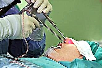
In the treatment of pathologies of the aortic valve, cardiac surgery is used:
- Prosthetics. The patient is fitted with a new valve - artificial or biological. Despite the fact that this operation normalizes hemodynamics best of all, there are also disadvantages: high risk of thrombotic complications; manipulation is contraindicated in old age.
- Balloon valvuloplasty - minimally invasive intervention used for stenosis. Description of the method: a special probe is inserted through the femoral artery, which expands the narrowed lumen.
- Balloon counterpulsation used in case of insufficiency. The technique of the method consists in the introduction of a catheter with an inflatable end. With the help of a tool, the affected dampers are leveled, as a result of which the latter begin to adhere tightly to each other.
Conclusions
The effective work of the aortic valve of the heart plays a significant role in maintaining adequate blood circulation parameters. Modern medicine offers a wide range of tools for effective surgical correction and drug support for patients with AK defects.