All substances necessary for the body are carried by the bloodstream. If failures occur in the cardiovascular system, then their transportation is disrupted. Among the diseases that affect this process, atrophy of the left ventricle of the heart stands out. It is a nutritional disorder of the tissues, as a result of which they cease to perform their functions. To fully understand the essence of the problem, it is necessary to find out why left ventricular atrophy of the heart is dangerous, what are its symptoms and how to treat this ailment at home.
The reasons for the development of the disease
To understand the features of left cardiac atrophy and to determine what it is, you need to look at the anatomy of the heart.

The heart is one of the most important human organs, whose functions include the blood supply to the entire body. It is based on the myocardium, which consists of striated muscle tissue. It allows the heart to contract, as a result of which blood is distilled between the atria and ventricles. In its structure, the myocardium resembles skeletal muscles, but, unlike it, the heart muscle does not obey the cerebral cortex. Signals to him are given by the vasomotor center, localized in the medulla oblongata.
The heart consists of 4 chambers, namely 2 atria and 2 two ventricles. Thanks to their alternating contractions, the blood circulates in a small and large circle, providing nutrition to all tissues of the body.
In general, the cardiac cycle looks like this:
- Systole (contraction) of the atria. They fill with blood and contract. From the right atrium, blood flow moves into the same ventricle, the same happens with the left.
- Contraction of the ventricles. The blood that has entered the left heart stomach is directed to the systemic circulation through the aorta. Contraction of the right ventricle leads it to the pulmonary artery (pulmonary circulation).
- Diastole (relaxation). This phase is the relaxation of all the chambers of the heart to fill the atria with blood. There is little oxygen in the bloodstream from the systemic circulation. This blood is called venous and enters the right atrium. The blood flow from the small circle is rich in oxygen, thanks to the pulmonary vessels. It is called arterial and travels to the left atrium.

The time it takes for the contraction phase is slightly longer than for relaxation. If everything goes well, then the person does not have problems with blood circulation. In the case of atrophy of the left ventricle of the heart, the body does not receive all the substances it needs due to the inability of the affected tissues to fully perform their functions. In this case, blood flow is disturbed, shortness of breath and swelling occurs. With the congenital form of pathology, developmental retardation is often observed.
Left ventricular atrophy may be congenital or acquired over time as an anomaly.
In the first case, the resulting dystrophy is a secondary phenomenon of myocardial cell disorder. Congenital heart defects and cardiomyopathy provoke its development. Anomalies in the structure of the heart muscle mainly affect the connective tissue of the myocardium and the valve component. Doctors still do not know much about cardiomyopathy, but they often eliminate the consequences of this disease, among which one can distinguish atrophy of the left ventricle.
The acquired form of the disease occurs in about 70% of patients. The reasons for its development are mainly as follows:
- Viral infections. Among them, influenza and the Coxsackie virus prevail. Left ventricular atrophy is a consequence of the effect of infection on the myosimplast. It is a fusion of cells, therefore it has several nuclei. After the attack of the virus, the myosimplast remains intact, but malfunctions occur at the genetic level, as a result of which dystrophic changes in the myocardium may begin. The degree of atrophy depends on the number of cells affected.
- Bacterial infections. Doctors distinguish scarlet fever from this group. This is due to the fact that the bacterium that causes the disease is very similar in its gene code to cardiomyocytes, which are the muscle cells of the heart. The immune system, after the defeat of the heart muscle by a bacterial infection, perceives its tissues as foreign and attacks them. This process leads to dystrophic changes in the myocardium.
- Lack of nutrition (ischemia) of the myocardium. In adults suffering from left ventricular atrophy, this is the main cause. This is usually associated with chronic ischemia, which gradually leads to degenerative changes in the heart muscle.
- Intoxication. Long-term exposure to toxins on the body negatively affects the heart muscle. The main culprit of intoxication is alcoholic beverages. Literally in 2-3 years of their intensive use, the myocardium undergoes significant atrophy.
Symptoms of pathology
Atrophy of the left ventricle of the heart can be detected by the clinical picture characteristic of this pathological process:
- Developmental retardation. The problem is inherent in the congenital form of the disease. The patient's tissues do not receive the required amount of the necessary substances for full-fledged growth, as a result of which developmental retardation is observed.
- Dyspnea. Atrophic changes in the left ventricle disrupt blood flow, which is why the body does not receive the required amount of oxygen. This phenomenon leads to ischemia of internal organs and tissues. To correct the situation, the respiratory center receives a signal to increase the amount of inhaled air. Shortness of breath is a consequence of this entire process and is designed to increase the concentration of oxyhemoglobin in the blood. It is a combination of hemoglobin and air in red blood cells.
- Swelling. They represent the second clear sign of acquired ventricular atrophy and speak of heart failure. Edema is manifested due to stagnation of venous blood, since the heart cannot fully perform its functions. Initially, the problem starts in the lower limbs. As the puffiness develops towards the top, it will be possible to conclude that heart failure is aggravated.
- Heart rhythm irregularities. Chronic nutritional deficiency leads to the death of cardiomyocytes. They perform not only the functions assigned to them, but also regulate the heart rate. With atrophic changes in the myocardium, the number of cardiomyocytes decreases, which leads to disruptions in the process of contraction of the heart.
Danger to the patient's life
Atrophy of the left ventricular tissue causes severe malfunctions associated with a lack of nutrition. The degree of danger to humans depends on the severity of dystrophic changes. Often this pathological process leads to such complications of the cardiovascular system as:
- atherosclerotic changes;
- fatal outcome;
- heart attack;
- angina pectoris;
- arrhythmia;
- pulmonary edema.
As the disease progresses, atrophied muscle areas are replaced by connective tissue, which is presented in the form of scars. Blood flow in this place is hampered and against this background the ability of the heart to contract weakens.
The course of therapy
Despite all the possibilities of modern medicine, doctors still do not know how to treat atrophy of the left stomach of the heart. The whole point of therapy is to stop pathological changes in the heart muscle.
There were also attempts to restore cardiomyocytes, thereby completely healing the patient. For this, the following drugs were used:
- "Preduct". It belongs to a group of medicines designed to relieve angina pectoris and improve myocardial metabolism in cardiac ischemia. The main active ingredient is trimetazidine dihydrochloride.Long-term intake of "Preductal" did not justify the hopes of scientists. The disease slowed down, but was not eliminated.
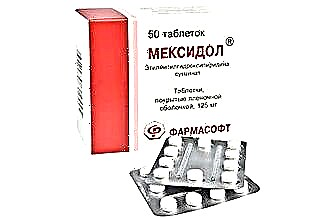
- "Mexidol" and "Riboxin". This combination is still not fully proven in the treatment of left ventricular atrophy. "Mexidol" is an antioxidant for ethylmethylhydroxypyridine succinate. Riboxin is intended to normalize myocardial metabolism and improve tissue nutrition. Its main active ingredient is inosine. Despite the promising description, many scientists consider their impact controversial and doubt the effectiveness of such a combination.
Due to the lack of actual research results, doctors try not to treat the disease, but to stop its development, stopping the consequences. Basically, the following groups of medicines are used:
- Diuretics They remove excess moisture from the body, reducing the stress on the heart muscle and solving the problem of edema.
- Beta-blockers. Drugs from this group inhibit beta-adrenergic receptors, thereby stabilizing heart contractions and reducing the degree of atrophy of cardiomyocytes.
- ACE inhibitors. Due to their effect, the production of angiotensin decreases, which constricts blood vessels, as a result of which the usual work of the heart is normalized. At the same time, the rate of development of atrophy decreases only in some patients.
- Calcium channel blockers and sartans. The above groups of medicines have been especially in demand in the treatment of left ventricular atrophy for the last 10 years. Calcium channel blockers prevent calcium from affecting the vessel walls, as a result of which they expand. Sartans are similar in effect to AFP inhibitors, but in this case angiotensin receptors are blocked.
Prophylaxis
It is extremely expensive and ineffective to treat left ventricular atrophy, so it is best to prevent it.
Prevention of the acquired form of the disease consists of the following rules:
- rejection of bad habits;
- playing sports;
- correction of the diet;
- full sleep;
- prevention of stressful situations;
- elimination of excess body weight.
Left cardiac atrophy is a severe and incurable pathological process. Due to its development, a person can experience various complications that threaten a fatal outcome. However, if you start a course of maintenance therapy in a timely manner, you can extend your life. To improve the effect, drug treatment must be combined with the observance of the rules of prevention.