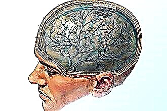
The human nose is surrounded by four pairs of air cavities, which perform part of the functions of the mucous membrane. The largest pair is located on the upper jaw to the right and left of the nose. The maxillary sinus is also called the maxillary sinus by the name of the British physician Nathaniel Highmore, who was the first to describe its main ailment - sinusitis.
Anatomical structure and physiological role of the maxillary cavities
 The maxillary sinuses are located inside the body of the upper jaw and have the shape of an irregular tetrahedral pyramid. The volume of each can vary from 10 to 18 cubic centimeters. The maxillary sinuses of the nose in one person can be of different sizes.
Inside, they are lined with a mucous membrane of ciliated columnar epithelium, the thickness of which is about 0.1 mm. The ciliated epithelium provides the movement of mucus in a circle to the medial corner, where the anastomosis of the maxillary sinus is located, which connects it to the middle nasal passage.
The structure of the maxillary sinuses is quite complex, in each of them 5 main walls are distinguished:
- The nasal (medial) one is most clinically important. Consists of a bone plate that gradually merges into the mucous membrane. It has an opening that provides a connection to the nasal passage.
- The front (front) is the most dense, covered with cheek tissues, it can be felt. Located in the so-called "canine (canine) fossa" between the lower edge of the orbit and the alveolar process of the jaw.
- The orbital (upper) is the thinnest, in its thickness there is a plexus of venous vessels and an infraorbital nerve, which can provoke complications in the lining of the brain and eyes.
- The posterior wall is thick, has access to the pterygopalatine node, the maxillary artery and the maxillary nerve.
- The lower wall (bottom) is the alveolar process, most often located at the level of the nose. If the bottom is lower, then the protrusion of the roots of the teeth into the walls of the maxillary sinus is possible.
The role of sinuses is not yet fully understood. Today, on the basis of the accumulated data, scientists distinguish the internal and external functions performed by them.
External functions include:
- secretory (providing mucus), protective, absorbing;
- resonator (participation in the formation of speech);
- reflex;
- participation in the olfactory process;
- regulation of intranasal pressure.
Also, the presence of cavities in the skull reduces the mass of the human upper jaw.
Internal functions include drainage and ventilation. Sinuses are capable of normal function only with constant drainage and aeration. The air flow passing through the passage forms air exchange in the sinuses, while the anatomy of the sinuses is such that at the moment of inhalation, air does not enter them.
Thus, in the maxillary sinuses, the structure is subordinated to the provision of nasal breathing. The reduced pressure in the voids during inhalation and the location of the anastomosis allow heated and humidified air from the sinuses to enter the inhaled air and warm it. On exhalation, due to a change in pressure, air enters the physiological voids, and their pneumatization occurs.
The ciliated epithelium, which covers each maxillary sinus from the inside, with the help of a strictly defined rhythmic movement of the cilia, move mucus, pus or foreign particles into the nasopharynx through the anastomosis. The length of the cilia is 5-7 microns, the speed is about 250 cycles per minute. At the same time, the mucus moves at a speed of 5 to 15 millimeters per minute.
The motor function of the ciliated epithelium depends on the pH level of the secretion (the norm is not higher than 7-8) and the air temperature (not lower than 17 degrees). When these indicators are exceeded, the activity of the cilia slows down. Violation of aeration and drainage leads to the occurrence of pathological processes in the sinuses.
An anastomosis is an oval or round hole about 5 mm long, covered with a mucous membrane with a small number of vessels and nerve endings. The cilia in the anastomosis constantly move the secret towards the exit. With normal cilia function and sufficient width, mucus does not accumulate in the sinuses, even in the presence of respiratory disease.
The diameter of the opening of the anastomosis is able to decrease and increase. The expansion is due to mild to moderate edema of the mucous membrane.
A constantly enlarged opening can cause the development of a cyst due to the ingress of a stream of air into the same point.
The prerequisites for the narrowing of the stroke can be as follows:
- severe edema due to a viral disease;
- the presence of polyps, tumors and various pathologies;
- congenital features of the human body (for example, a naturally narrow notch).
The narrowed passage does not provide a quick drainage of mucus that stagnates inside. In this case, inflammation begins, pathogenic microbes multiply rapidly and pus is formed, which indicates the development of sinusitis.
The reasons for the development of sinusitis (sinusitis)
Sinusitis is an inflammation of the maxillary accessory cavities, most often due to an infection that has entered them through the blood or during breathing. However, the causes of the onset of the disease can be identified much more.
The main ones are:
- untreated or poorly treated rhinitis (runny nose);
- infection of the nasopharynx with pathogenic bacteria and viruses;
- past illnesses (ARVI, flu), running cold;
- injury to the wall of the maxillary sinus;
- long-term stay in a room with warm and dry air, as well as in a chemically hazardous production;
- poor oral hygiene, especially teeth;
- hypothermia of the body, drafts;
- weakened immunity;
- violation of the secretory function of the glands;
- disturbed anatomy (curvature) of the nasal septum;
- overgrowth of polyps and adenoids;
- allergic reactions;
- severe ailments (neoplasms, mucosal fungus, tuberculosis).
A prerequisite for the development of sinusitis is often the long-term use by the patient of drops with a vasoconstrictor effect, intended for the treatment of the common cold.
Symptoms and types of disease
Depending on the localization of the inflammatory process, sinusitis can be right-sided, left-sided or bilateral. The patient's condition gradually deteriorates, especially in the evening. The main signs of the disease:
- discharge from the nasal passages in which mucus and pus are present;
- a feeling of pressure in the bridge of the nose, aggravated by tilting the head;
- nasal congestion, complete or alternating left and right sides;
- memory impairment and poor sleep;
- high temperature in acute form (up to 39-40 degrees), chills;
- malaise, weakness, lethargy, fatigue, a sharp decrease in performance;
- pain in the nose, passing to the forehead, temples, eye sockets, gums, eventually covers the entire head;
- labored breathing;
- voice changes (nasal).
With sinusitis, a profuse nasal discharge is most often observed. This is due to the accumulation of mucus, blood clots and pus in the nasal cavities. Depending on the color of the discharge, experts distinguish between the main stages of the development of the disease:
- white - the initial stage or stage of recovery (with a thick consistency);
- green - the presence of acute inflammation in the sinuses;
- yellow - there is pus in secret, this is an acute form of the disease that requires the intervention of an otolaryngologist.
The most difficult is the situation in which there are secret blood clots and streaks. The maxillary sinuses are located near vital organs, therefore, with an advanced disease, serious complications are possible.
Depending on the cause of the disease, the following types of sinusitis are distinguished:
- Rhinogenic occurs after poorly treated viral infections, influenza, rhinitis. Most
 a common type of sinusitis (over 60% of all cases).
 - Polyposis is caused by the growth of polyps in the nasal passage, as a result of which the natural anatomy of the cavity is disrupted and congestion develops.
- Allergic appears against the background of the influence of aggressive external factors, causing a strong response of the body, mainly has a seasonal character with exacerbations in the spring and autumn months.
- Odontogenic manifests itself against the background of inflammatory processes in the accessory cavities caused by staphylococci, streptococci, Escherichia coli. A common cause is dental disease and poor oral hygiene.
 a common type of sinusitis (over 60% of all cases).
Diagnosis and treatment of sinusitis
To determine the causes and stage of development of the disease, the otolaryngologist examines the nasal passages. To obtain a more complete clinical picture, fluoroscopy or computed tomography of the cavities is performed.
With conservative therapy of sinusitis, general and local methods are combined, aimed at suppressing the pathogenic microflora, cleansing and sanitizing the organ:
- Drops and sprays. They give a vasoconstrictor effect (Galazolin, Naphtizin, Xylometazoline), may also contain excipients with antihistaminic properties (Vibrocil, Cetirizine) or local antibiotics (Bioparox, Polydex).
- Antiseptics in the form of drops and rinsing solutions ensure the outflow of secretions and cleansing of the nasal passages (Miramistin, Dioxidin, Protorgol, Furacilin, Chlorhexidine). It is necessary to listen to the doctor's recommendations, since many of them have contraindications for children or pregnant women.
- Antibiotics The most commonly used drugs are the penicillin group (Flemoklav, Amoxiclav), cephalosporins (Cefixim, Pantsef), macrolides (Clarithromycin, Azithromycin).
If drug treatment does not give the desired effect or the anastomosis is completely blocked, the doctor may resort to puncturing the sinus wall.

During puncture, the accumulated exudate is pumped out with a syringe, the cavity is washed and anti-inflammatory drugs and antibiotics are injected into it. The puncture can be cured in a shorter time. Also in modern medicine, special YAMIK catheters and the method of balloon sinusoplasty are used to avoid puncture. Spy software for modern smartphones for tracking a subscriber or listening to conversations - how to find and install. Instagram spy apps and special programs on smartphones are increasingly used by us in our everyday life.
Untimely treatment of sinusitis can lead to serious complications - meningitis, inflammation of the optic nerve, osteomyelitis of the facial bones
Cleansing the sinuses at home
Additional to the drug therapy can be the use of alternative methods of treatment. You can cleanse the affected cavities using the following recipes:
- Rinsing with a solution of sea salt (no more than 1 teaspoon per half liter of boiled water). With your head tilted, you should pour the solution into the nostril using a teapot or syringe without a needle, without creating strong pressure. Water should flow out through the other nostril.
- After rinsing, it is recommended to drop 2 drops of thuja essential oil into each nostril. This procedure must be repeated three times a day for two weeks.
- 20% alcoholic tincture of propolis is mixed with vegetable oil (1: 1) and instilled into each nostril.
- Sea buckthorn oil is dripped into the nostrils or used for inhalation (10 drops per pot of boiling water, breathe for 10-15 minutes).