The external nose of man is a unique creation of nature, absent from other living beings on earth. It has the shape of an irregular triangular pyramid, and its frame is made up of the nasal bones and cartilage of the nose (two-thirds). This organ not only performs physiological functions, but also gives a person an individual unique look.
The structure of the outer area of the sense organ
 The external nose (in Latin nasus externus) is anatomically subdivided into two sections: bone and cartilaginous. The pyramid of the nose in its rear part has no edge, since it passes into the nasal cavity.
The other two facets covered with skin, connecting at an acute angle, form the back (dorsum nasi), which extends from the root to the top of the organ, and below it is connected to the nasal wings (alae nasi).
The back is formed by the facial bones, septal and lateral cartilages, and the wings are formed by the small and large cartilages of the wings.
- Lateral cartilage (cartilago nasi lateralis) paired, similar to a triangle of irregular shape. With its posterior edge, it adjoins the anterior segment of the nasal bone, the upper segment of the inner edge - to the same cartilage from the opposite side, with which it can sometimes grow together, and the lower segment - to the nasal bridge. At the bottom, it reaches the lateral pedicle of the large wing cartilage.
- Large wing cartilage (cartilago alaris major) is also paired. In a circle, it surrounds the nostril (nares), which form the input into the nasal cavity. Includes lateral (crus laterale) and medial (crus mediale) legs. The medial, between which the nasal plate is inserted, separate the nostrils. The lateral pedicles are longer and wider than the medial ones; they form the wing frame. Also, 2-3 small cartilages (cartilagines alares minores) can adjoin the lateral legs in the upper posterior region, adding rigidity to the structure.
- Sometimes in the tissues of the sensory organ of honey, the main ones are additional cartilages of various sizes and shapes, called sesamoid.
Each nasal cartilage is connected to the others, as well as to the osteostructures by fibrous tissue and is covered by the perichondrium.
A special place in the structure of the external nose has a nasal septum (septum nasi), which divides the internal space of the organ into two relatively equal parts.
 In its structure, two areas are distinguished: lower (mobile) and upper (bone). The larger segment of the movable region is the cartilage of the nasal septum, which looks like a quadrangular plate of irregular shape. Its postero-upper part rests against the angle between the opener and the ethmoid plate. The antero-inferior edge reaches the medial legs and wedges between them, is the most mobile in the nose. In children, the entire septum is membranous; over time, part of it hardens. The movable region of the scaffold can move under the influence of the muscles that surround the nasal wings.
Cartilage pain
Very often, patients who feel pain in the nasal frame turn to otolaryngologists and traumatologists. The reasons for their occurrence may be the following factors:
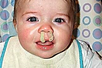
- Injuries (bruises and fractures) of the external nose are very common, causing pain in the tip and back, especially on palpation. In this case, the work of the respiratory system is disrupted due to displacement, damage or bifurcation of the frame. Bleeding (in people suffering from hypertension, blood and liver diseases), suppuration (with stagnation of the secreted blood in the tissues), swelling are possible. It is necessary to urgently go to the hospital, since there is a threat of the development of an abscess and the complete destruction of the cartilaginous tissue.
- Damage to the soft tissues of the sensory organ. It is typical for people involved in sports, especially in contact forms. However, the reasons can also be everyday occurrences: falls, bruises, traffic accidents.
- Sinusitis. The inflammatory process and the accumulation of pus in the maxillary sinuses leads to the fact that pain is given to different parts of the skull.
- Rhinitis. The defeat of the mucous membrane provokes congestion, fever, general malaise.
- Neoplasms. In the initial phases, external changes are invisible, however, bleeding, purulent discharge, and shortness of breath are observed. In a neglected form, a change in the shape of external tissues becomes obvious.
- Boils, purulent acne can give off pain in all the component parts of the organ due to the presence of a large number of nerve endings in the soft integument.
 Damage to the bony part of the nose is capable of transmitting pain to the movable part, both with and without displacement.
 Damage to the bony part of the nose is capable of transmitting pain to the movable part, both with and without displacement.
In case of injury to the nasal septum, it is necessary to apply any cold compress to the affected area (ice, meat from the freezer, a bottle of cold water). If bleeding occurs, cotton swabs can be gently inserted into the nostrils. After that, be sure to contact a surgeon or a trauma center. It is strictly forbidden to feel or try to straighten damaged parts yourself. This can lead to displacement of soft tissues, complication of treatment and a serious cosmetic disadvantage.
Curvature of the nasal septum
Curvature of the nasal septum can occur for physiological, traumatic and compensatory (age) reasons. Its symptoms are:
- Difficulty in nasal breathing (from partial blockage of the stroke to complete cessation of air intake). Depending on the shape of the curvature, there may be a problem with breathing in the right or left way. Often, the patient does not feel this, since the nasal cavity adapts and compensates for the deficiency at the expense of other structures. In older people, the passage of air is difficult due to the depletion of the body's compensatory resources.
- Snoring at night.
- Drying out inside the cavity.
- Changing the shape of the organ. There is a shift to the right or left after fractures, dislocations. Without treatment, parts of the frame may not heal properly.
- Allergies. If breathing is impaired, allergic reactions can continue for a longer period, despite the treatment. Itching is felt inside, secretion is secreted.
- Inflammation of the paranasal sinuses (frontal sinusitis, sinusitis, ethmoiditis) in a chronic form. In this case, polyps may appear on the mucous membrane.
The consequences of curvature and lack of oxygen can be changes in the vascular system, blood, genital area, weakening of the immune system and exposure to adverse environmental influences. The otolaryngologist performs an examination using an endoscope or a mirror, determining the degree of curvature and studying the structural features of the patient's cavity. Urine and blood tests are also done.
 The most effective method of eliminating the curvature of the septum today is surgery. There are several types of operative correction of lintel deformation:
- Septoplasty is an intervention to correct the septal plate, often combined with a submucosal vasotomy.
- Lateral conchopexy is an operation to bring the turbinates closer to the lateral wall to expand the narrowed passages.
- Cristotomy - removal of the ridge of the septum that interferes with breathing.
- Vasotomy - removal of the spongy (cavernous) tissue of the inferior shells to reduce them when filled with blood in the event of the development of vasomotor rhinitis.
- Rhinoplasty is the correction of aesthetic deficiencies of the sensory organ, often carried out in parallel with septoplasty.
The most common septal surgery is septoplasty. Modern technologies make it possible to make an incision inside the cavity, so that the scar is invisible. The cartilage exfoliates from the perichondrium and mucous membrane, after which it is separated from the bone and displaced in the desired direction. Then the spines and ridges are removed.The deformed cartilage is modeled and put in place, after which it takes its natural physiological position due to its biomechanical properties. In this case, the bridge continues to perform its natural support function.
The operation lasts no more than an hour, the postoperative period can last up to 2 weeks.
Methods for performing rhinoplasty
Rhinoplasty is an operation that aims to change the shape and size of the outer nose. The reasons for the correction are most often post-traumatic and congenital deformities. In addition, many people, especially women, turn to specialists to improve the appearance of their senses.
The operation can be performed both on the mobile and on the bone sections. The surgeon is able to shorten, raise or narrow the tip, straighten the back, and remove the hump.
In case of asymmetry or large width, intervention on the nasal wings is necessary. There are three main types of rhinoplasty:
 Open (American school) - incisions are made on the post dividing the nostrils and incisions are made in the cavity, the skin is separated from the frame and wrapped. Visually examining the operating field, the surgeon takes the necessary actions. After the end of the intervention, sutures are applied for 5 days, and self-absorbable threads are used inside. Tampons are inserted into the nostrils, a fixing (plaster) bandage is applied on top.
- Closed (European school). All incisions are made in the nasal cavity and are not visible. There are no external scars. The stages of correction are mainly carried out blindly. This method is more often practiced with small adjustments. Plaster fixation is used in the postoperative period.
- "Non-surgical". This is a change in the shape or shape of an organ using subcutaneous injections of fat or other fillers. Most often, hyaluronic acid is used for this. This procedure does not injure the patient, so there is no need to carry it out in a hospital setting.
 Open (American school) - incisions are made on the post dividing the nostrils and incisions are made in the cavity, the skin is separated from the frame and wrapped. Visually examining the operating field, the surgeon takes the necessary actions. After the end of the intervention, sutures are applied for 5 days, and self-absorbable threads are used inside. Tampons are inserted into the nostrils, a fixing (plaster) bandage is applied on top.
Rhinoplasty can be performed only after the formation of the nasal skeleton, i.e. not earlier than 18 years old. The optimal age for this kind of surgery is 25-35 years. At a later age (after 45-50 years), doctors are reluctant to perform rhinoplasty, since the risk of complications increases, and tissues regenerate more slowly.