In the face of the skull there are cavities - voids called the paranasal sinuses. They function as resonators, thanks to which the mass of the head bones is reduced. Each nasal sinus communicates with the nasal cavity through an anastomosis - a narrow connecting passage. There are several types of paranasal, or paranasal sinuses, differing from each other in location, size, structure.
Common to all paranasal sinuses
The anatomy of the nose and paranasal sinuses is especially actively formed during the first 5 years of life. Together with the nasal cavity, the paranasal sinuses form a single functional system.

All paranasal sinuses have walls that are dotted with numerous holes. Connective tissue cords, nerves, and blood vessels pass through these holes. However, through the same holes in the cavity can penetrate:
- pus,
- toxins,
- pathogenic flora,
- cancer cells spreading to the orbital region, pterygopalatine fossa, etc.
Due to the fact that the structure and physiology of the nose and paranasal sinuses allows the possibility of the traffic of pathogens, the development of secondary diseases and the emergence of complications after, at first glance, a harmless infection of an individual sinus is often observed.
Functions
One of the main tasks of the sinuses is to ensure the safety of the brain, eye sockets, facial nerves, arteries and veins. The anatomy of the paranasal sinuses normally suggests the possibility of unhindered removal of constantly produced mucus, the physiological function of which is to neutralize pathogens. The mucus is discharged through the fistulas, which must be open for this, and moves to the exit due to the ciliated epithelium, covered with many cilia.
With the onset of a cold, mucus production increases.
However, in the case of significant edema of the mucous membrane and blockade of the anastomosis, exudate accumulates in the cavities. This could be due to:
- an infection leading to swelling of the mucous membranes,
- the structure of the form of the anastomosis, where the main role is played by their narrow diameter,
- curvature of the septum,
 the appearance of a polyp, tumor.
- hypertrophy of the membrane.
 the appearance of a polyp, tumor.
In addition to the protective function, a distinction is made between:
- resonator, thanks to which an individual tone of voice is formed,
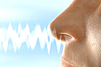
- respiratory (in the process of nasal breathing, air circulates freely through the nasal passages, moistens and warms up),
- olfactory (the task is performed thanks to the smell-recognizing epithelial tissue).
Anatomical abnormalities
The paranasal sinuses are diverse, and their number and shape may vary from person to person. So, for example, according to statistics, the frontal sinuses are generally absent in 5% of people. In addition, topographic relationships may be disturbed, thickening or thinning of the walls of bone tissue, on the surface of which there may also be congenital defects. Such abnormalities occur in the late phase of prenatal (intrauterine) development.
Common anatomical abnormalities include asymmetry in the frontal and maxillary sinuses. And to rare - the complete absence of the maxillary cavity and the division of the maxillary sinuses in half by a bony septum.

This division can occur both vertically (front and back) and horizontally (top and bottom).
Cracking of the upper wall of the maxillary sinus, which communicates with the lower orbital canal or the cavity of the orbit, is more common. The concavity of the front wall, combined with the extension of the nasal wall into the lumen of the sinus, threatens the penetration of the needle under the cheek when trying to puncture.
Anatomy and physiology also depend on a genetic factor that can cause deformation of the facial and brain skeletons, as well as on metabolism.
For all sinuses in the paranasal region, the presence of slit passages of communication with the surrounding formations (dehiscence) is considered abnormal. For example, due to the occurrence of dehiscences:
- ethmoid labyrinth sometimes communicates with the frontal and sphenoid sinuses, orbit, cranial fossa;
- the slit in the lateral wall of the main sinus contributes to the contact of its mucosa with the dura mater (cerebral) of the middle cranial fossa, the pterygo-palatine fossa, the superior orbital fissure and the optic nerve, the cavernous sinus and the internal carotid artery;
- thinning of the wall of the sphenoid sinus can lead to contact with the abducens and block nerves, with the branches of the oculomotor and trigeminal nerves.
Maxillary (maxillary) sinuses
Paired caves that are located in the thickness of the bone. In an adult, the volume of each can reach 30 cm3 (max), but the average volume is about 10 cm3... In volume form, it resembles a three-sided pyramid. Its three walls stand out:
- The upper (orbital) is the thinnest of the three, which is especially noticeable in its posterior part. Often, it is in these places that cracks appear, and sometimes bone tissue is completely absent. Inside the wall, from the infraorbital foramen, passes the canal of the inferior orbital nerve. If the channel is absent, the nerve and accompanying blood vessels are attached to the mucous membrane. However, in the event of inflammatory processes with such an arrangement, the likelihood of intraorbital and intracranial complications increases.
- The lower (cavernous bottom) is located next to the posterior part of the alveolar process (that is, near the upper jaw), therefore it sometimes happens that the sinus is separated from the four posterior upper teeth only by soft tissues. This proximity increases the risk of sinus inflammation due to odontogenic injury.
- The inner wall (it is also the lateral wall of the nasal cavity) - normally corresponds to the middle and most of the lower nasal passages. In the posterior region of the semilunar notch under the middle part of the nasal concha, the maxillary sinus opens through this wall with an opening into the nasal cavity. Everywhere, except for the lower sections, this wall is thin enough so that a therapeutic puncture can be made through it.
Paired maxillary sinuses often differ in volume, while both shells (right and left) have bays (small additional depressions): alveolar, palatine, zygomatic, frontal.

Frontal (frontal) sinuses
They are paired cavities that are located in the thickness of the frontal bone, namely, between the plates of the scales and the orbital part. The right and left shells are usually separated by a thin septum. However, due to the peculiarities of the formation, options are possible when:
- the septum is displaced to the left or right, which sometimes causes a significant difference in the size of the shells,
- the septum may have openings that communicate with each other the frontal sinuses,
- cavities may be missing on one or both sides,
- the sinus can extend to the frontal scales, as well as to the base of the skull, together with the perforated plate of the ethmoid bone.
The frontal sinus communicates with the concha through the frontal-nasal canal. Its outlet is located in front of the mid-nasal passage.
Frontal shells become a continuation of the anterior cells of the ethmoid labyrinth, therefore, in the case of inflammation of one formation, the infection often spreads to another.
- The anterior wall is the place through which the sinus is punctured or opened. The orbital nerve exits through the supraorbital notch.
- The lower wall is the thinnest of all, which makes it easier for infection to enter the orbit from the frontal concha.
- The cerebral wall, through which the infection can penetrate into the anterocranial fossa, separates the shells from the frontal lobes.
Lattice maze

A collection of thin-walled cells, consisting of bone tissue. Their average number is about 7-8, but the number can vary from 2 to 15. The cells are located in 3-4 rows, with a conditional division into front, back and middle. They are located in the unpaired symmetrical ethmoid bone - in the notch of the frontal bone. The posterior cells are in contact with the canal through which the optic nerve passes (sometimes it passes directly through them). Often, the lattice labyrinth reaches the most distant cavities of the facial skeleton, bordering on vital organs.
The mucous membrane of the labyrinth is innervated from the nasal ciliary nerve - a branch of the orbital nerve. In this regard, many diseases occurring with the defeat of the trellis labyrinth are accompanied by pain. Due to the fact that the olfactory filaments pass in the narrow canals of the bone ethmoid plate, during the development of edema due to compression, olfactory disorders are not uncommon.
Sphenoid (main) sinus
 Due to its location in the sphenoid bone (behind the ethmoid labyrinth above the vault of the nasopharynx and choanas), the main sinus has a second name - the sphenoid. In an adult, this sinus is divided into right and left non-communicating parts, which in most cases do not coincide in size and have independent exits into the nasal passage. In total, five walls of the cavity are described:
- Front. It consists of two parts: nasal and trellis, which corresponds to the posterior cells of the trellis labyrinth. The thinnest anterior wall smoothly passes into the lower one, turning into the nasal cavity. There are small rounded holes in it, through which the main sinus communicates with the nasopharynx. They are located at the end of the upper nasal concha.
- Back. A frontally located wall, less than a millimeter thick (with large volumes of the sinus), which causes the risk of damage to it during operations.
- Upper. Corresponds to the bottom of the sella turcica, in which the intersection of the optic nerve (enveloped by the arachnoid) and the pituitary gland are located. In the case of inflammation of the sphenoid sinus, it often passes to adjacent formations, sometimes affecting the olfactory tract or even the anteromedial surface of the frontal lobes of the brain.
- Lower. Thick (about 12 mm) wall corresponding to the fornix of the nasopharynx.
- Side. These walls border directly on the neurovascular bundles, which are located on the sides of the sella turcica. They can both absorb the optic nerve canal and come into contact with it. An infection can enter these formations through the wall at the border with the cavernous sinus and optic nerve.
Along with the listed sinuses, mention should be made of the pterygopalatine fossa located behind the tubercle of the lower jaw. Its clinical significance is great, since if the nerves located in the fossa are involved in the inflammatory process, neuralgic syndromes of the facial part occur.
Sinus inflammation: types and symptoms
 Depending on which sinus the inflammatory process takes place in, there are:
- sphenoiditis - inflammation affects the sphenoid sinus,
- sinusitis - the maxillary cavity is affected,
- frontal sinusitis - the frontal zones are involved,
- ethmoiditis - the process takes place in the cells of the lattice labyrinth.
Inflammation of the mucous membrane can affect one or more sinuses at once. This inflammatory process takes place in different forms:
- acute form with pronounced symptoms,
- recurrent - with less pronounced repetition of signs of acute inflammation,
- chronic.
The chronic form of the course of the inflammatory process, which often concerns the maxillary and slightly less often the frontal sinuses, lasts about 2-3 months, even if therapeutic measures are used. Signs of a chronic process include:
- Discharge from the nose of a purulent, mucous, watery or mixed consistency.
- Difficulty breathing due to blockage of the nasal passages.
- Sore throat and reflex cough arising from swelling of mucous masses along the back of the pharynx.
- Headaches mainly in the nose, forehead and eyes.
- Violation of the olfactory function.
- The growth of polyps from the paranasal sinuses into the nasal passages.
Unlike children, adults are more likely to experience a viral infection of the nasal mucosa, which spreads to the sinuses. Less commonly, blood diseases and dental conditions are the cause. The odontogenic factor is essential in the defeat of the maxillary sinuses. A bacterial factor, most often in the form of staphylococci, can join and activate a viral infection against the background of the "busy" immune system.
Normally, microorganisms and microparticles when inhaled, passing through the nasal cavity along with air, enter the caves of the sinuses, where the ciliated epithelium captures them and neutralizes them with the formation of mucus excreted outside. This mechanism can be disrupted by the curvature of various bone formations with anatomical deformation of the shells, as well as unfavorable factors affecting the protective properties of the epithelium: dry air, tobacco smoke, chemical burns, tissue atrophy and necrosis, a suppressed state of the immune system, etc. as well as a consequence of an allergic reaction.
 Among the most common common symptoms of sinus inflammation are:
- runny nose with thick greenish discharge and pus,
- headache, which increases with pressure drops, when the head bends, pressure on the area in the area of the nasal sinuses, as well as a feeling of fullness in these areas,
- condition of nasal congestion,
- an increase in body temperature up to 38C,
- morning and night cough.
Due to congestion, a person begins to breathe through his mouth, speaks in nasal voices. At the same time, an unpleasant odor is often felt from the mouth.
With sinusitis, headaches associated with a pathological increase in intracranial pressure are one of the main symptoms. Pain in the forehead and sinuses can be pulsating or compressive, which is characteristic, first of all, for an acute form. In addition to the above signs, it is noted:
- decreased sense of smell (or loss of it),
- lacrimation and fear of light,
- sometimes - swelling of the upper eyelid or cheek.
In the chronic course of the disease, discharge flows down the wall of the pharynx, provoking a night cough. In the mornings and evenings, there is a characteristic pain radiating to the region of the eye sockets. When pressing on the inner corner of the eyes, the pain spreads to the entire face.
Treatment of inflammation
Treatment of inflammation is carried out by conservative or surgical methods, depending on the indications. Conservative methods involve the removal of edema of the mucous membrane, the destruction of pathogens, the creation of conditions for the drainage of mucus and the organization of the patency of the mouth of the sinus.
In the treatment of acute forms without the need to remove cysts, polyps, eliminate septal curvatures, use:
- vasoconstrictor drugs - to relieve edema,
- local antibiotics - for purulent inflammation,
- antiseptic solutions in combination with rinsing through a puncture of the most convenient and thin wall,
- oil preparations for moisturizing dry mucous membranes, eliminating crust,
- saline solutions when washing to moisturize and normalize the drainage of exudate.
Method "Cuckoos" for sinusitis
 Washing is used only if there are no violations in the structure of the anastomoses, provided that the fluid circulates normally through the nasal cavity. It is performed without anesthesia. The patient lies on his back. A catheter is inserted into one nostril to deliver medication, and a tube with a vacuum pump is inserted into the other nostril to pump fluid.During the procedure, the patient pronounces the onomatopoeic "cuckoo", which gave the name to the method, to prevent the medicine from entering the airways through the throat. When the medication is delivered, a slight pressure is created to facilitate the flushing of the exudate. In the treatment of sinusitis, 5 sessions are usually prescribed.
Sometimes washing is combined with laser treatment, which is used to relieve swelling.
Sinus catheter flushing
Sinusitis can be treated without a puncture with the help of Yamik. For washing, catheters are introduced to the patient, through which high and low pressure is created (for this, an air balloon is connected). Through one catheter, the contents of the sinuses are pumped out, and a medicinal solution is supplied through the other. The procedure is performed under local anesthesia.
Cyst
The cyst is detected by x-ray. Without it, patients hardly notice the neoplasm until it reaches a significant size, comparable to the volume of the sinus. In this case, symptoms characteristic of sinusitis begin to appear: headaches, a feeling of fullness, difficulty with nasal breathing. A cyst occurs when the ducts of the mucous gland are disturbed, due to which mucus is collected in a spherical capsule. It can be eliminated only surgically after determining its exact location using CT and MRI:
- The classical method involves an incision in the wall under the upper lip, which is associated with prolonged scarring and frequent subsequent recurrence of sinusitis.
- The endoscopic method is performed using an endoscope with a camera through the anastomosis, which excludes traumatic complications.
Fungal infection
 Fungal inflammation is not considered rare. The fungus affects one sinus or several at once.
At risk are HIV-infected and diabetics, and the likelihood of infection increases in people:
- conducting topical steroid treatment,
- taking antibiotics regularly
- using drug therapy that suppresses the immune system,
- who have undergone radio and chemotherapy in connection with an oncological disease.
The inflammatory reaction is most often provoked by fungi of the genera Candida, Mukor, Aspergillus, Rhizopus.
In this case, the symptoms of a fungal infection are similar to a bacterial infection. The picture of the course of the disease can vary from slow development to rapid growth of fungal formations with severe manifestations. Again, we note that this system will not guarantee a win, but will allow you to stop in time, saving your money, without "going into a minus". Has the necessary certificates in the public domain, the casino is legally operating play fortuna the best online casino livejournal rudy1977. The unique official casino website https://pinupcasino-play.online/ has many slot machines. But this is not the case, each slot has a moment of recoil, which must be hit. Wild is a special bonus symbol, the essence of which is to replace pictures with the ones necessary for a winning combination. An accurate diagnosis is established first with the help of radiological images, and then it is clarified by conducting histological and mycological analyzes. In the case of fungal infection, antifungal drug treatment is most often combined with surgical intervention aimed at removing polyps from the sinus.
Features of inflammation in children
90% of all cases of sinus inflammation in children are bacterial in nature. Due to the fact that at this age there are a large number of variants of manifestations, sometimes difficulties with diagnosis arise. With inflammation in newborns, the diagnosis is based on:
- cough,
- smell from the mouth,
- switching to mouth breathing,
- blocked nasal passages.
A specific symptom can be attributed to swelling of the eyelids and / or displacement towards the eyeball, which is associated with the location of the ethmoid sinus near the eye sockets, which in infants are separated from the sinus by an incompletely formed wall. These manifestations are observed against the background of general symptoms: loss of appetite, tearfulness, worsening sleep. Older children may additionally complain of pain and swelling around the eyes. They also have nasal congestion, alternating with purulent-mucous discharge.