Difficulty breathing, snoring, runny nose, bleeding and pain in the nose are problems that bother a large number of people, but rarely become a reason to see a doctor. In most cases, the cause of these symptoms is a curvature of the nasal septum. About 80% of people on the planet suffer from this physiological anomaly to one degree or another. In most cases, minor deviations from the norm do not disrupt the normal functioning of the nose and do not require surgery, but if the above symptoms become chronic and create constant discomfort, then surgery becomes inevitable.
The structure and function of the nasal septum
 The nasal septum is a plate located in the nasal cavity and dividing it into two approximately equal parts. In the depths of the nose, it consists of a thin bone, and in the front part it is made of cartilaginous tissue. The area of cartilage is softer and protrudes forward (you can touch it by running your hand along the midline of the nose), which makes it very vulnerable. Inside, both sides of the septum are covered by a mucous membrane.
Thanks to this bone-cartilaginous plate, the inhaled air is divided into two streams and moves into the respiratory tract.
Here it is ensured uniform warming, cleansing and moisturizing. Thus, any disturbances in the structure of this part of the respiratory system lead to malfunctions in its functioning and can lead to unpleasant consequences (inflammation, swelling of the mucous membranes, snoring during sleep, headaches, disruption of the heart and nervous systems, etc.) ).
For example, when inhaling, in a person with a curvature, the wing of the nose can stick to the septum and accordingly block the access to air. In this case, the patient usually begins to breathe through the mouth, and this, in turn, leads to drying out of the mucous membranes. In addition, sinus aeration is impaired. The frontal and maxillary (maxillary) sinuses do not receive the necessary air exchange. As a result, the outflow of mucus becomes difficult, and inflammatory processes begin, the consequence of which can be chronic sinusitis, sinusitis, tonsillitis, etc. Moreover, breathing through the mouth can provoke oxygen starvation of the brain, which affects the mental abilities of a person.
Signs of curvature
Most people do not even suspect that they live with a deformation of the osteochondral plate in the nose, since this does not interfere with the work of their respiratory system, which, with minor deviations from the norm, adapts and provides air exchange in the required volume. However, if you find the symptoms below, it is recommended to consult an otolaryngologist and surgeon. since in such cases the problem must be eliminated promptly:
- trouble breathing through the nose;
- visual change in the shape of the nose;
- mouth breathing;
- nosebleeds;
- dry nose;
- decreased olfactory ability;
- chronic rhinitis;
- frequent respiratory infections;
- snoring while sleeping.

Causes and types of curvature
In most cases, curvature of the septum occurs during adolescence and adolescence (13-18 years), although cases of congenital physiological abnormalities are also known. One of the most common causes of pathology is physiological. In this case, there is a discrepancy between the growth rates of the cartilaginous and bony parts of the septum. Sometimes the size of the nasal cavity becomes insufficient to accommodate the separating plate, and it begins to bend.
Deformation can also be caused by injury (dislocation, nose fracture). In this case, the bones of the nose are first displaced, and then they do not heal properly.
In addition, compensatory curvature is distinguished, which appears due to the influence of irritating factors (polyps, tumors, foreign bodies on the nasal mucosa) and hypertrophy - the uneven development of one of the nasal concha.
There are several types of deformities of the nasal septum. Depending on the shape, S-shaped and C-shaped curvatures are distinguished. On the septum, ridges, spines, thickenings can also form, and a dislocation of the quadrangular cartilage is possible. In addition, there are 3 degrees of severity of the deformation of the nasal septum:
- slight deviation from the midline (I degree);
- the protruding section of the osteochondral plate is located between the midline and the lateral wall of the nose (II degree);
- the protruding portion of the osteochondral plate practically touches the lateral wall of the nose (grade III).
Diagnosis and surgery
 By itself, a slight deformation of the dividing plate is not a reason for surgery. It is enough to observe the hygiene of the nasal cavity, avoid staying in dusty places, try not to overcool and complete the treatment of respiratory diseases to the end.
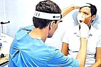
However, if your nasal septum hurts or at least one of the above symptoms is observed, then at least you need to contact a specialist so that they make a diagnosis based on the examination. As a rule, otolaryngologists conduct an examination using a rhinoscope.
In addition, MRI, CT and X-ray are effective examination methods.
Nowadays, there are several techniques for correcting the curvature of the nasal septum. If the deformity is not too large and affects only the cartilaginous part, which, moreover, has not been broken, you can resort to laser correction. This operation is performed under local anesthesia. With the help of a laser, the doctor heats up the areas of cartilage tissue that need to be removed. After completing this procedure, the nose is fixed in an even position with two gauze swabs inserted into the nasal passages.
A more common method of surgical intervention is septoplasty, which is performed using both modern endoscopic techniques and traditional surgical techniques.
This operation is usually performed after the completion of the formation of the osteochondral plate, starting at the age of 18. The duration of the operation is on average 1-2 hours. The surgeon makes a small incision in the mucous membrane and peels it off where it is necessary to remove the deformed portion of cartilage or bone. After that, the mucous membrane is returned to its place, and the septum is fixed with gauze tampons.
For several days after the operation, the patient is forced to breathe through the mouth, since the nasal cavity remains sealed. During this time, changes in ambient temperature should be avoided. Also, the patient is prescribed a course of antibiotics to prevent the development of infections and pain relievers to relieve pain. After 7-10 days, the septum should no longer hurt, but since the swelling of the mucous membrane may not completely disappear, some difficulty in breathing through the nasal cavity is possible. Return to normal life usually occurs 2 weeks after surgery. As with any operation, doctors recommend that patients avoid serious physical exertion and temperature changes for a month.