An aneurysm is a bulge or distension of the wall of a blood vessel. This disease can develop not only in the aorta, but in all arteries of the body, including the carotid arteries. An aneurysm in the carotid artery can gradually stretch and weaken the structure of the vascular wall, as a result of which, when the pressure rises, it ruptures. This can occur both in the extracranial (extracranial) region of the carotid artery - in the neck, and in the intracranial, which is located in the brain. Rupture of an aneurysm of the carotid artery is a very dangerous complication of this disease, since it entails multiple ischemic and metabolic changes in the brain.
Symptoms and clinic of the condition
 The course of this disease can be asymptomatic, with small protrusions, or gradually increase due to structural changes in the aneurysmal wall. Symptoms of a carotid artery aneurysm include:
- Dizziness;
- constant ringing in the ears;
- unreasonable headaches;
- feeling of chronic fatigue;
- sleep disorders.
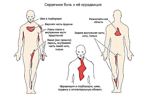
The larger the size of the aneurysmal formation, the brighter the manifestations of symptoms become: the headache becomes more frequent and intensifies, discomfort appears in the chest region, shortness of breath, and a decrease in visual acuity. There may also be hoarseness of the voice, impaired coordination, a feeling of vascular pulsation carried out to the head.
Symptoms of aneurysmal protrusion include a picture of a mini-stroke or transient ischemic attack (TIA). A TIA is an episode of poor circulation in the brain with temporary stroke-like symptoms that may include mild facial disturbances, excessive fatigue or sleep, muscle weakness on one side of the body, slurred speech or difficulty speaking, and dizziness. TIA is often considered a warning sign that a true stroke could occur in the future if something is not done to prevent it.
 Carotid aneurysms can form blood clots in arteries that block blood flow to the brain. According to a study published by the University of Maryland Medical Center (UMMC), carotid thrombosis was present in 6.2% of the total number of participants screened. Carotid artery thrombosis can lead to frequent strokes that cause paralysis, brain damage, or death. The blood clot can also break away from the aneurysm and travel to the brain, which subsequently leads to a blockage of the cerebral artery.
A severe headache that starts suddenly is a characteristic sign of a ruptured aneurysm in the carotid artery. The pain is so intense that most people describe it as "the worst pain ever felt." An extremely severe headache is usually accompanied by nausea and vomiting, neck numbness, and, in some cases, temporary loss of consciousness.
Secondary symptoms caused by an aneurysm of the carotid artery arise from its pressure on the surrounding structures. When an aneurysm expands, nerves and veins are compressed, resulting in symptoms such as facial swelling and tingling, numbness in the face or mouth, loss of voice or hoarseness, difficulty speaking, and difficulty swallowing.
An unruptured carotid aneurysm can cause vision problems. It can cause blurred or double vision, chronically dilated pupils, and pain radiating to the forehead. If the carotid artery ruptures, temporary loss of vision may also occur.
Diagnostics: how to recognize and distinguish a disease?
It is necessary to differentiate this disease with the following pathologies: ischemic stroke, cavernous sinus syndrome, cluster headache, fibromuscular dysplasia, neurofibromatosis, cervical form of lymphogranulomatosis.
The diagnostic algorithm for carotid artery aneurysm includes:
 anamnestic data;
- Physical examination findings (palpable sac-shaped swelling in the neck)
- Ultrasound of the neck. This study serves to determine the size and expansion of the aneurysm;
- angiography - the gold standard for diagnosing carotid aneurysm;
- contrast CT - has such advantages as easy and fast applicability; is a minimally invasive method that makes it possible to see changes in the vascular wall, with a volume of less than 1 mm;
- Magnetic resonance angiography is a non-invasive technique that can visualize vascular structures without the need for contrast media or X-rays. MRA can show thrombosed portions of the aneurysm and residual blood flow characteristics.
 anamnestic data;
Treatment and rehabilitation of a patient with carotid artery aneurysm
The goal of aneurysm treatment is to reduce symptoms and reduce the risk of complications. Before choosing any method, it is important to discuss the potential benefits, risks, and side effects.
If the aneurysm is small and does not cause any symptoms, expectant management is chosen with careful monitoring of the condition, examination with ultrasound, CT or MRI every six to twelve months.
If there is a risk of complications, surgical treatment is used. Surgical intervention entails resection of that part of the carotid artery, which is associated with the aneurysm, and replacement of the removed area with a graft.
 Another option for surgical treatment is the placement of an endovascular stent. The operation is performed depending on the size of the aneurysm and its location relative to the rest of the branches of the carotid artery. The surgeon goes through a puncture in the femoral artery using catheters to guide and deliver the stent graft to the site of the aneurysm. X-ray guidance is used to place a graft made of artificial material in the region of the protruding vessel wall. The stent is then expanded inside the artery and held in place with metal hooks rather than sutures. The advantage of this treatment method is to reduce the operational risk and shorten the patient's rehabilitation time.
Rehabilitation of patients consists of blood pressure control, smoking cessation, adherence to a diet (reduction in the diet of fats, carbohydrates), taking medications to thin the blood, and moderate physical exertion.
Conclusions
So, aneurysm of the carotid artery is a really rare disease that should be taken into account when conducting differential diagnostics of diseases with the presence of a pulsating formation in the neck and neurological symptoms. Such patients may have complaints such as dysphagia, headache, neck pain, a feeling of retro-orbital pressure, otalgia, and symptoms of cardiovascular disease. Early diagnosis and treatment is essential as cerebrovascular complications and death occur in 50 to 70 percent of cases. But the satisfactory results of surgical treatment convince the need for aggressive therapy of this disease.