Atheroma of the ear is a benign neoplasm that occurs on the human skin in places where the sebaceous glands accumulate when their excretory ducts are blocked. According to statistics, this problem is relevant for 5-10% of the population, mainly for middle-aged people. The most common atheroma of the auricle is on the earlobe, since it contains mainly adipose tissue. A cyst behind the ear is diagnosed in only 0.2% of cases of neoplasms in the facial area.
Reasons for the appearance

The main reasons for blockage of the sebaceous gland ducts are hormonal problems (excessive production of hormones) and metabolic disorders. However, many factors are known that can also contribute to the formation of a cyst (atheroma) in the ear:
- seborrhea of the scalp;
- simple and phlegmous acne;
- increased sweating;
- malfunctions of the endocrine system;
- diabetes;
- sloppy piercings and head injuries;
- increased testosterone production;
- prolonged exposure to the sun or hypothermia;
- non-compliance with hygiene rules;
- prolonged stay in polluted rooms.
Under the influence of one or more reasons, the duct of the gland narrows, the secret becomes thicker and is not able to stand out. Over time, a cystic cavity (capsule) appears in the place of the plug, where detritus (cholesterol crystals, epithelial cells, frozen fat) gradually accumulates. It is first felt with the fingers, and then becomes visible to the eye.
Symptoms
The development of atheroma in the ear for a long time (several months) is asymptomatic. The person does not feel any discomfort or pain. However, in the process of development of education at an early stage, the following main symptoms of the disease appear:
- small size and round shape, resembling a ball;
- on palpation, it is defined as a dense formation that can move under the skin;
- the skin above the ball cannot be folded;
- consists of a capsule with a curdled secret inside.
A small inconspicuous neoplasm may not interfere with a person at all, but the risk of a worsening of the clinical picture is still sufficient great.
In the case of inflammation of atheroma behind the ear or in the earlobe, the symptoms change:
- the ball increases and becomes clearly visible;
- itching and burning sensation appears;
- a subcutaneous abscess develops (pain, redness and an increase in skin temperature over the induration).
A blocked gland can open up on its own. In the best case, a wound is formed, which, with minimal care and disinfection, will quickly heal. In the worst case, pus may leak out, but detritus will begin to accumulate in the remaining capsule. In addition, it is possible to attach a secondary infection with the manifestation of headache, fever, nausea, weakness, fatigue. It should be noted that the cyst is not able to transform into an oncological process and provoke cancer.
Sometimes inflammation of the sebaceous gland behind the ear is confused with another benign neoplasm - a lipoma. The main distinguishing feature is the presence of a partial adhesion to the skin and a small outlet of a dark color (or white in case of suppuration) in the cyst.
Diagnostics
The basis of diagnosis is a primary visual examination by a specialist (otolaryngologist or surgeon), palpation with fingers to pinpoint the location of the formation and identify the location of the blocked duct.
However, to distinguish it from other neoplasms, such as hygroma, fibroma or lipoma, the doctor may advise to undergo a morphological or histological examination. Based on their results, it can be concluded that there are no signs of malignancy in the tumor.
Treatment of the disease
Modern medicine recognizes only one really effective way to treat this benign formation of the auditory organ - surgery. Due to the specific structure of the formation and the presence of a hard capsule, it is impossible to get rid of it only by methods of conservative therapy. Conservative methods are used in parallel with surgical ones:
- for preliminary removal of severe inflammation that interferes with the operation;
- in the postoperative period to accelerate recovery and prevent wound infection.
Since there is no chance that the seal will dissolve by itself, it will sooner or later have to be removed.
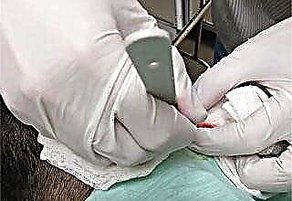
There are three main ways to do this today. All of them are performed under local anesthesia.
- Laser. It is used in cases where there is no inflammatory process. This usually occurs in the early stages of the disease. The following techniques are used:
- Photocoagulation (evaporation) is carried out when the size of the formation is up to 5 mm. As a result, a crust remains at the site of the operation, after the natural removal of which, after 1-2 weeks, there is no scar.
- Laser excision. An incision is made in the skin with a conventional scalpel, the shell is lifted so that the border between the capsule and the surrounding tissues is clearly visible. Then the cells of the adhesion of the membrane to the skin are evaporated with a laser beam, after which the entire cyst is removed with forceps, a drainage is introduced into the wound and sutured. The stitches are removed after 10 days. The method is used for neoplasms with a diameter of 5-20 mm.
- Laser vaporization of the capsule. It is used for large neoplasms. Through a deep incision, all detritus is removed, after which the dense cells of the epidermis, which form a shell, are evaporated by a laser. Wounds heal for about two weeks, the scar is hardly noticeable.
- Radio wave. It is used only in the absence of suppuration and with a small ball size. The cells of the formation are killed in a strictly delineated area, as a result of which the cyst disappears. it
 the only method that gives 100% results. It does not injure the tissue, and the stitches are not applied.
 - Traditional surgical. Most often it is carried out in a polyclinic, with the exception of cases with severe suppuration, which are treated in a hospital. A scalpel over the capsule is made an incision in the skin (or two incisions near its base), the capsule is removed together with the shell. If the integrity of the shell is violated, then the purulent contents are first removed, and then the shell is selected piece by piece. A particle of the capsule remaining in the wound can lead to a relapse of the disease (according to statistics, such cases are not more than 3%). After the operation with a scalpel, a rather noticeable scar remains, which can only be reduced by laser resurfacing.
 the only method that gives 100% results. It does not injure the tissue, and the stitches are not applied.
After surgery, care for the wound consists in washing it with hydrogen peroxide, applying Levomekol ointment and gluing it with a plaster or medical glue (usually 2-3 weeks).
Attempts to squeeze out the cyst on their own will not lead to success, since the shell remaining inside with the cells that produce the secret will again fill up with sebum after a while. In addition, during the squeezing process, the surrounding tissue can be damaged or infected through micro-damage to the skin. If the atheroma is located behind the ear, home treatment is very dangerous, since large blood vessels and lymph nodes pass in this area of the head.
Traditional methods
A person's fear of an operation makes him look for other ways to cure the disease. With atheroma of the earlobe, treatment with folk remedies is used most often. Several popular recipes:
- Melt mutton fat, cool to body temperature, rub into the affected area at least 5 times a day. You can add a little crushed garlic and vegetable oil to the composition.
- Squeeze out the aloe juice and apply to the desired place 2-3 times a day.
- Boil a hard-boiled chicken egg, peel.Then remove a thin film from the egg and apply to the seal. Repeat for several days.
- Bake the onion in the oven, then knead until mushy and mix with shavings of laundry soap. Apply to the bump and secure with a bandage or plaster.
These funds can only be used with the permission of a doctor and on a non-inflamed cyst.