The eardrum is a thin (0.1 mm) plate of connective tissue that separates the middle and outer ear. If the eardrum is retracted, the causes should be sought in the inflammatory processes of the auditory (Eustachian) tube, which connects the tympanic cavity and the nasopharynx. Both children and adults are susceptible to this disease.
Causes of occurrence

The Eustachian tube is a canal that balances the pressure inside the ear with atmospheric pressure, which is a prerequisite for the normal functioning of the hearing aid. Since the width of the passage is only 2 mm, then any inflammatory process of its walls blocks the passage, disrupts the drainage function and triggers catarrhal inflammation. This disease is called eustachitis or tubo-otitis and can be acute or chronic.
The main cause of acute tubo-otitis is the spread of infection from the upper respiratory tract and nasopharynx into the mucous membranes of the auditory tube in such diseases:
- angina;
- flu;
- ARVI;
- pharyngitis or rhinitis;
- whooping cough;
- measles;
- scarlet fever;
- Infectious mononucleosis.
The causative agents of the disease are staphylococci, streptococci and viruses, as well as pneumococci in children.
Less commonly, it is caused by fungal infection, allergies (hay fever, allergic rhinitis) and specific microflora (syphilis, tuberculosis).
The development of chronic eustachitis is due to the presence of inflammatory processes in the nasopharynx, which are permanent:
- chronic sinusitis and rhinitis;
- tonsillitis;
- adenoids.
Complicated air passage due to curvature of the nasal septum or benign neoplasms in the pharynx and nasal cavity (polyps, adenoids, scars, tumors) also contributes to the onset of the disease.
Development and main symptoms
Due to a violation of the patency of the Eustachian tube (partial or complete), there is a violation or cessation of its ventilation. The retracted tympanic membrane indicates that the air remaining in the internal cavity has already been absorbed, and the pressure in it is reduced. This leads to the fact that a transudate with fibrin and protein (yellowish or greenish in color) is drawn into the cavity. It complicates the movement of the ossicles and membrane, and leads to a decrease in hearing up to a third of the norm. Later, neutrophils and lymphocytes can enter the cavity, which can provoke inflammation.
Such processes become prerequisites for the catarrhal form of otitis media with the risk of becoming purulent, especially in people with reduced immunity. This is fraught with the appearance of adhesions (adhesive otitis media), a sharp deterioration in hearing and the need for a complex operation or hearing aid.
The main symptoms of tubo-otitis can be bilateral or manifest in one ear:
- hearing loss;
- heaviness in the head;
- ear congestion;
- autophony (the echo of your own voice) and tinnitus;
- fluid transfusion is often felt;
- limescale and salt deposits;
- thinning of the lintel.
Changes in air pressure inside the cavity lead to painful sensations, a feeling of pressure and distention in the ear. The patient does not have any other negative sensations and fever. Sometimes, when you yawn or swallow saliva, your hearing improves for a while.
The reason for this is an increase in the lumen of the tube with the contraction of the corresponding muscles.
The acute form of the disease can turn into a chronic one, the features of which are periods of exacerbation and remission. At the same time, the diameter of the pipe is steadily decreasing, which leads to sticking of its walls and the presence of constant symptoms of eustachitis.
Diagnostics
The diagnosis is established based on the medical history and additional examinations, in particular:
- otoscopy and microotoscopy (visual inspection using special systems);
- audiometry (determining the level of hearing loss at certain frequencies);
- acoustic impedance measurement (detecting the compliance of the jumper to determine the presence of liquid behind it);
- research using a tuning fork.
With otoscopy, a sharply distinguished process of the malleus is noted, as well as a retracted tympanic membrane, the causes and treatment of which are determined by objective and subjective methods.
Subjective ways:
- Sample of an empty throat. The patient takes a deep breath of air.
- Toynbee's test. The same, but with pinched nostrils.
- Valsalvi test. A deep breath is taken, the mouth is closed, the nose is pinched, and an exhalation is made.
Objective method - blowing through the Eustachian tube and measuring the result obtained by means of audiometry and otoscopy. If, after blowing, hearing improves and the retraction of the membrane weakens, then the cause of the problems is in the ear canal.
In addition, a throat swab is taken to identify the pathogenic microflora and determine the antibiotics necessary to combat it.
Treatment of the disease
When treating Eustachitis, complex therapeutic measures are carried out, which include several areas:
- Elimination of the primary source of the disease, which caused the violation of the patency of the auditory tube:
 antibiotic therapy;
- tonsillectomy, removal of adenoids;
- correction of the nasal septum;
- removal of tumors;
- restoration of full nasal breathing.
- Removal of puffiness, inflammation or allergic reaction:
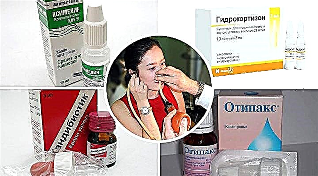
- vasoconstrictor drugs in the nose (vibrocil, sanorin, nasol, nasivin);
- oral antihistamines (desloratadine, suprastin, claritin).
- Hearing restoration and prevention of hearing loss development:
- the introduction of a solution of epinephrine or hydrocortisone using a catheter into the tympanic cavity;
- pneumatic massage;
- physiotherapy (UFO, UHF, laser therapy, electrical muscle stimulation).
 antibiotic therapy;
To avoid getting infected mucus from the nasopharynx into the ear, the patient is not recommended to blow his nose strongly. Allocations must be removed without stress.
If conservative help did not produce the desired effect, then a puncture of the membrane and drainage of the cavity is done. In more severe cases, bypass surgery is performed. The tympanic cavity is washed through the shunt.
Pneumatic massage as part of therapy
Pneumatic massage is usually used at the catarrhal stage of the disease or at the stage of recovery. It can be done in a hospital or at home.
In otolaryngological departments, the following are used:
- apparatus "APMU-Compressor", which presses on the jumper with the help of baro impulses;
- Politzer balloon (rubber bulb with a tube), where the impact is carried out by manual air pressure.
At home, after the instructions of the ENT doctor, patients perform manual pneumomassage. The most common techniques are:
- Close the ears tightly and press lightly on the shells, creating air pressure. Perform 10 presses 1-3 times a day. escortnavi
- After a deep exhalation, insert the index fingers into the ear canals, gently move them, and then pull them out sharply.
- After taking a deep breath, pinch your nostrils and close your mouth. Try to exhale through the nose with an effort, then swallow air. This is a homemade option for blowing out the ear canal.