A malignant tumor of the middle ear, auditory canal or auricle - ear cancer - is manifested by the formation of a node, bleeding granulations, ulcers. These early signs of ear cancer are most often accompanied by itching, noise, pain, and discharge. One-sided hearing impairment often occurs. However, the presence of certain symptoms depends on the type of tumor. In the case of the spread of a malignant formation, signs of damage to the cranial nerves appear. The enlargement of regional lymph nodes is associated with lymphogenous metastasis.
Cancer classifications and symptoms by type
 Statistics show that ear cancer accounts for about one percent of the total number of cancers.
There is no gender difference: men and women are equally at risk of the disease. Most often these are people after 40 years. Localization statistics have the following picture:
- 80% - ear cancer,
- 15% - tumor of the external auditory canal,
- 5% - a neoplasm in the middle ear.
There is a classification of ear neoplasms according to the criteria of localization, causality, type of growth, microscopic and histological structures. Based on the localization criterion, the disease can affect the middle ear and the external one, with a division into malignant neoplasms of the auricle and a tumor of the external auditory canal.
The causality factor allows us to divide this oncology into:
- primary, when benign cells are transformed into malignant ones,
- secondary, when the focus of growth is in nearby organs (for example, the nasopharynx), followed by germination in the area of the hearing organs.
By the type of growth, exophytic tumors are distinguished, which grow in the lumen of the organ, and endophytic, growing mainly in depth. Histological examination reveals several types of lesions that develop at different rates and external differences:
- Spinocellular epithelioma (squamous cell carcinoma, squamous cell carcinoma). A fast-growing malignant tumor growing from epithelial cells (flat cells located in the surface layer of the epidermis).
- In the auricle it looks like a wart with a wide base and an outgrowth that bleeds slightly.

- The ear canal resembles a kidney-shaped outgrowth or erosion that sometimes spreads throughout the ear canal.
- In the auricle it looks like a wart with a wide base and an outgrowth that bleeds slightly.

In 75% of cases, it affects the skin of the face and scalp. This type of epithelioma accounts for about 25% of all cancers of the skin and mucous membranes. More susceptible to the disease are people of the Caucasian race with fair skin, who quickly burn in the sun. More often it affects people over 60 years old, and in children, predominantly genetically determined tumors are recorded.
In 98% of cases, it metastasizes by the lymphogenous pathway. Reaching the lymphatic system, cancer cells spread through the lymphatic vessels and, lingering in the local lymph nodes, begin to last. Hematogenous distribution with blood flow occurs in only 2% of cases.
- Basalioma (basal cell carcinoma). Most often occurs on the skin of the face and neck. Basalioma is able to slowly grow both into the subcutaneous tissue and into the nearby bone and muscle tissues, but it either does not give metastases at all, or metastasis occurs at later stages.
At the initial stages, basal cell carcinoma may look like a normal pimple that slowly grows in size without causing painful sensations. A grayish crust forms in the center of the pimple. If removed, a temporary indentation remains on the skin, which soon becomes crusty again. A specific sign of this neoplasm is the presence of a dense ridge around the tumor. The roller itself consists of fine-grained formations that resemble miniature pearls, and becomes especially noticeable when the skin is stretched. Due to the expansion of superficial vessels, the presence of spider veins is often noted.
If there are several nodular lesions, they may merge over time.
With the germination of basal cell carcinoma into the surrounding tissues, a pronounced pain syndrome occurs. Depending on the form of the disease, symptoms may vary:
- Nodular-ulcerative is manifested by a supracutaneous rounded nodular-like seal, which increases over time, acquiring an irregular configuration.
- The warty form is characterized by the appearance of a hemispherical dense nodule resembling a cauliflower.
- The large-nodular (nodular) form is a single supracutaneous node with vascular asterisks on the surface, growing outward.
- The pigmented form, due to the pigmented center or periphery, may resemble melanoma, but this type of basalioma is characterized by a specific “pearl” ridge.
- Sclerodermiform formation differs from others in that, as it grows, it turns into a dense, flat plaque with a rough surface and clear contours.
- The cicatricial-atrophic form is characterized by the formation of an ulcer in the center of the nodule, which spreads to the edge. At the same time, the scarring process begins in the center, which creates a characteristic picture with a pronounced edge and a scar in the center.

The disease develops more often in people after 40 years. Prolonged exposure to direct sunlight directly contributes to its occurrence. At risk are light-skinned people, as well as those in contact with carcinogens and toxic substances.
- Sarcoma. A very rare neoplasm in this localization, which arises from the underlying "immature" connective tissue. Depending on the affected tissue, chondrosarcoma (cartilage), osteosarcoma (bone), myosarcoma (muscle), liposarcoma (fatty) are distinguished. It is characterized by slow growth in the area of the auricle and rapid growth in the ear canal. Sarcomas, unlike other cancers, are not "tied" to specific organs.
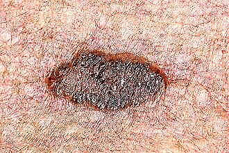
- Melanoma. Rapidly developing, but rare disease for this localization. Malignant neoplasm occurs due to atypical degeneration of melanocytes - pigment cells. The occurrence of this pathology in people with dark skin is unlikely, however, in the statistics of diseases, a family predisposition can also be traced.
The varied shape, consistency and color of melanoma makes it difficult to self-diagnose by the appearance of the formation. Melanoma can be of almost any shape and contain inclusions of several colors ranging from gray-black and brown to blue and pink-purple. There are depigmented formations. More often than elastic, melanoma has a dense texture. A characteristic feature of melanoma is the absence of skin formation on the surface. External manifestations depend on the form of the disease:
- Superficially spreading when it appears, it looks like a black or brown non-convex pigment spot measuring 5 mm or less. The period of lack of growth above the skin surface can last up to 7 years. The vertical phase is accompanied by a sharp activation of the oncological process.
- The nodular form is also similar to a mushroom or a polyp. The color varies between blue-red and black. Despite the vertical structure of the formation, a horizontal phase of growth is also recorded in the process.
- Lentigo-melanoma in the horizontal phase can last 10-20 years. With the transition to the vertical phase, the lesion focus becomes uneven in color, and the edges become uneven.
The division of the oncological process into 4 stages is used in clinical practice to determine the type of cancer spread.
- In the first stage, there is a lesion of the skin of the outer ear and the mucous membrane of the middle.
- In the second stage, the underlying cartilage of the outer ear and the bone structures of the middle within the compact layer are affected.
- The third stage is characterized by going beyond the boundaries of the compact layer with the defeat of regional lymph nodes.
- The tumor affects adjacent anatomical structures, conglomerates of infected lymph nodes are formed, which include deep cervical lymph nodes.
Specificity of malignant neoplasms of the hearing organs
 The asymptomatic initial stage of cancer of the outer ear is replaced by noticeable itching, noise, and localized pain. An ulcer, granulation, or nodule that affects the ear canal bleeds more often than a mass that affects the ear. In addition to blood, mucus or pus may be released.
Hearing impairment begins with germination into the internal sections, which is accompanied by:
- increased pain intensity,
- paralysis of facial muscles (with damage to the facial nerve),
- intracranial complications.
In terms of symptoms, middle ear cancer in the initial stages resembles chronic otitis media. As characteristic signs, suppuration and hearing loss are more common than others.
Granulations appear in the ear canal, the growth of which gradually becomes more active and is accompanied by bleeding. Hearing impairment progresses. The pain syndrome increases with the germination of the underlying tissues (while the pain radiates to the neck and temple).
In the case of involvement of nearby anatomical structures in the oncological process, symptoms of their defeat occur: sharp pains with the involvement of nerves (trigeminal, facial), muscle paralysis, impaired swallowing and motor function of the lower jaw. The movement of the tumor towards the brain is characterized by the development of carcinomatous meningitis. The "affected" internal carotid artery begins to bleed profusely.
Diagnosis and treatment options
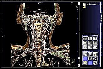
The diagnosis is made on the basis of otoscopy, radiography (with a tumor of the middle ear), histological and cytological studies. The direction and volume of the area affected by the tumor is determined using MRI and CT of the brain. In the case of external ear disease, oncology is distinguished from tuberculoma, weeping eczema, lupus erythematosus, ear inflammation and benign neoplasms. With cancer of the middle ear, it is differentiated with osteomyelitis of the temporal bone, chronic purulent otitis media, tuberculosis, syphilis, carotid chemodectoma.
Treatment and prognosis depend on the neglect of the disease and the level of involvement of adjacent anatomical structures in the pathological process:
- In the first stage, patients are prescribed radiotherapy. After a course of radiation therapy and in the presence of cancer residues, electroexcision is performed.

- At the next stage, a combination therapy is shown: preoperative radiotherapy and surgical removal of the formation or, in case of damage to the external auditory canal, the entire auricle. In some cases, the electroresection of the node may be an option for classic surgery.
- In the later stages, radiation therapy is used, followed by excision along with the fiber of pathological lymph nodes.

In the case of limiting the growth of a neoplasm to the area of the tympanic cavity after a radical operation, a cure is possible. When the bones (zygomatic and main), middle cranial fossa, carotid artery and meningeal membranes are affected, the prognosis is poor.