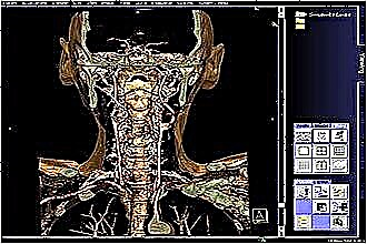
A complete examination is required to make a diagnosis of a laryngeal lesion. It includes a doctor's examination, an analysis of anamnestic information, on the basis of which additional laboratory and instrumental research is prescribed. The most informative diagnostic method is considered to be an MRI of the larynx, but the examination is also carried out using X-rays and an endoscopic method (direct laryngoscopy).
Benefits of MRI
 Due to its high information content, non-invasiveness, and painlessness, the study is widespread in medical practice. The procedure provides the maximum amount of information about the state of soft tissues, blood vessels, lymph nodes, cartilage structures. It is possible to increase the information content with the help of intravenous contrasting, which more clearly visualizes oncological, cystic formations.
Computed tomography of the larynx is prescribed by an otolaryngologist, oncologist, surgeon to determine the treatment tactics of a conservative or surgical direction.
Among the symptoms, when a tomography is prescribed, it is worth highlighting:
- shortness of breath, swallowing;
- hoarseness of voice;
- neck deformity that is visually noticeable;
- soreness when palpating;
- nasal congestion in the absence of sinusitis, which indicates the possible presence of a Thornwald cyst;
- headaches, dizziness;
- swelling of soft tissues.
Thanks to MRI of the throat, the following pathological conditions and diseases are diagnosed:
- the consequences of injuries in the form of cicatricial changes;
- the presence of a foreign body;
- inflammatory foci, lymphadenitis;
- abscess, phlegmon;
- cystic formations;
- oncological diseases.
In addition, the study of the larynx with a tomograph makes it possible to trace the dynamics of the progression of the disease, to evaluate the effect of the treatment, including in the postoperative period.
The high resolution of the tomograph makes it possible to identify an oncological focus at the initial stage of development
The advantages of an MRI of the throat are:
 harmlessness, since the study is carried out using a magnetic field;
- non-invasiveness, which does not imply a violation of the integrity of tissues, penetration into hollow organs;
- painlessness;
- high information content with the possibility of 3D image reconstruction;
- the ability to differentiate between benign and malignant neoplasms.
Limitations in the use of MRI are associated with the high cost and the need to study bone structures, when MRI is not so informative.
No preparation for diagnostics is required. Before starting the examination, it is necessary to remove jewelry containing metal. For 6 hours before the study, it is forbidden to eat if the use of contrast is expected.
Among the contraindications to MRI of the throat, it is worth noting:
- the presence of a pacemaker;
- metal prostheses;
- metal fragments in the body;
- pregnancy (1) trimester.
In the presence of metal elements in the human body, when exposed to a magnetic field, they can move somewhat from their place. This increases the risk of injury to the surrounding structures and tissues.
Features of laryngoscopy
Laryngoscopy refers to diagnostic techniques that make it possible to examine the larynx, vocal cords. There are several types of research:
- indirect. Diagnostics is carried out in a doctor's office. A small speculum is located in the oropharynx. With the help of a reflector and a lamp, a beam of light hits the mirror in the mouth and illuminates the larynx. Today, such laryngoscopy is practically not used, since it is significantly inferior in informational content to the endoscopic method.
- Direct - performed using a flexible or rigid fibrolaryngoscope. The latter is often used during surgery.
Indications for laryngoscopy include:
- hoarseness of voice;
- pain in the oropharynx;
- difficulty swallowing;
- feeling of a foreign object;
- an admixture of blood in sputum.
The method allows you to establish the cause of the narrowing of the larynx, as well as assess the degree of damage after injury. Direct laryngoscopy (fibroscopy) is usually done to remove foreign objects, take biopsy material, or remove polyps.
Indirect laryngoscopy is performed on an empty stomach to avoid aspiration (ingestion of gastric contents into the airways). Removable dentures are also required.
Direct endoscopy of the larynx is performed under general anesthesia, on an empty stomach, after collecting some information from the patient, namely:
- the presence of allergic reactions;
- regular medication intake;
- cardiac diseases;
- violation of blood clotting;
- pregnancy.
Contraindications include
- ulcerative lesion of the oral cavity, epiglottis, oropharynx due to the high risk of bleeding;
- severe cardiac, respiratory failure;
- severe swelling of the neck;
- laryngeal stenosis, bronchospasm;
- uncontrolled hypertension.
An indirect examination is carried out in a sitting position. The patient opens his mouth, the tongue is held in place with a napkin or fixed with a spatula.
To suppress the gag reflex, the doctor irrigates the mucous membrane of the oropharynx with an anesthetic solution.
A small mirror is located in the oropharynx, after which the examination of the larynx and ligaments begins. A beam of light is reflected from a refractor (a mirror fixed on the doctor's forehead), then from a mirror in the oral cavity, after which the larynx is illuminated. To visualize the vocal cords, the patient needs to pronounce the sound "A".
Direct endoscopic examination is performed under general anesthesia in an operating room. After the patient falls asleep, a rigid laryngoscope with a lighting device at the end is inserted into the oral cavity. The doctor has the opportunity to examine the oropharynx, ligaments, or remove a foreign body.
When conducting a direct examination, while maintaining the patient's consciousness, the mucous membrane of the oropharynx should be irrigated with an anesthetic, a vasoconstrictor is instilled into the nasal passages. After that, the flexible laryngoscope is advanced along the nasal passage.
The duration of the procedure takes about half an hour, after which it is not recommended to eat, drink, cough or gargle for two hours. This will prevent laryngospasm and choking.
 If, during laryngoscopy, surgery was performed in the form of removal of a polyp, it is necessary to follow the doctor's recommendations for the management of the postoperative period.
After laryngoscopy, you may experience nausea, difficulty swallowing, or hoarseness.
When conducting a biopsy, an admixture of blood in the saliva may appear after the study.
The risk of complications after examination increases with obstruction of the respiratory tract by tumor formation, polyp, in case of inflammation of the epiglottis. After a biopsy, bleeding, infection, or damage to the respiratory tract may occur.
According to the results of the study, the doctor can diagnose inflammatory diseases, detect and remove a foreign body, assess the severity of traumatic injury, and take a biopsy if an oncological process is suspected.
X-ray in the diagnosis of diseases of the larynx
For the diagnosis of throat pathologies in otolaryngology, ultrasound and tomography are most often used.Despite the availability of modern instrumental examination methods, an X-ray of the larynx is also used, although it is not a highly informative technique.
Usually, x-rays are performed in patients who cannot use laryngoscopy. X-ray diagnostics does not require preparation. X-rays are taken straight, lateral, and anterior and posterior.
Taking into account the need to obtain an image in a certain projection, the patient is placed on his side or chest. The research is carried out as follows:
- an X-ray tube generates a beam beam;
- radiation passes through tissues of different density, as a result of which more or less dark shadows are visualized in the image.
Muscles pass the radiation flux well. Bones, having a high density, block their path, which is why the rays do not appear on the film. The more X-rays hit the picture, the more intense their shadow coloration.
Hollow structures are characterized by a black shade. Bones with low radiological throughput are displayed in white on the image. Soft tissues are projected with a gray shade of varying intensity. According to indications, contrasting is used, which increases the information content of the method. The contrast agent in the form of a spray is sprayed onto the mucous membrane of the oropharynx.
The picture assesses the X-ray anatomy of the larynx. When viewed from the side view, many anatomical structures can be seen, such as the root of the tongue, the body of the hyoid bone, the epiglottis, the ligamentous apparatus (vocal, epiglottis-arytenoid), the ventricular fold, the vestibule of the larynx, as well as the ventricles of Morgagni and the pharynx, localized behind the larynx.
High-quality radiography of the larynx allows the doctor to assess the diameter of the lumen of the hollow organs, the glottis, the motor ability of the ligaments, and the epiglottis.
Cartilaginous structures reflect radiation poorly, therefore they are practically not visualized in the image. They begin to appear during calcification, when calcium is deposited in tissues.
At the age of 16-18, calcification occurs in the thyroid cartilage, then in the rest of the laryngeal cartilage. By the age of 80, complete calcification of the cartilaginous structures is noted.
Thanks to the X-ray, displacement of the organ, a change in its shape, and a decrease in the lumen are diagnosed. In addition, foreign bodies, cystic formations, oncopathology of benign or malignant origin are visualized.
Among the indications should be highlighted:
- traumatic injury;
- tracheal stenosis with diphtheria;
- chemical, thermal burn;
- violation of the movement of the vocal cords.
Contraindications include pregnancy, however, when using protective equipment, research may be allowed.
Based on the clinical picture, the doctor determines which methods of examining the larynx will be the most informative in this case. Thanks to a comprehensive examination, it is possible to diagnose pathology at an early stage of development. This makes it possible to choose the optimal therapeutic course and achieve complete recovery.

 harmlessness, since the study is carried out using a magnetic field;

