Middle ear diseases are the most common form of hearing ailments. They affect adults and especially children. To date, doctors have developed a large number of modern techniques that can provide treatment for the middle ear, the symptoms and treatment of the most common diseases of this localization will be discussed below.
Acute otitis media
 This middle ear disease occurs in two main forms: catarrhal and purulent.
In the catarrhal form, the tympanic cavity, the mastoid process and the auditory tube are affected. The main pathogens are bacteria (pneumococci, streptococci, staphylococci). The development of the disease is also facilitated by:
- infectious diseases;
- hypothermia;
- diabetes;
- avitaminosis;
- kidney disease.
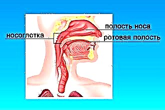
The penetration of pathogenic microflora occurs mainly through the auditory tube from the nasal cavity in case of diseases of the mucous membrane (influenza, ARVI, acute respiratory infections, rhinitis).
This is facilitated by improper blowing of your nose (through two nostrils at the same time), sneezing, and coughing.
In childhood, infection is easier due to the structural features of the pipe (it is wide and short). Also, there are frequent cases of infection through the blood with scarlet fever, measles, tuberculosis. The growths of the adenoids that overlap the mouths of the auditory tubes often lead to relapses and the transition to a chronic form.
Symptoms characteristic of this middle ear disease are:
- severe pain (aching or throbbing), radiating to the temporal and occipital region of the head;
- feeling of stuffiness and noise;
- hearing loss;
- temperature increase;
- deterioration in sleep and appetite;
- eardrum reddened and painful to touch.
Treatment is usually carried out at home and bed rest is prescribed. Hospitalization is carried out only with signs of complications (meningitis, mastoiditis). Conservative treatment of catarrhal otitis media is carried out as follows:
- Removal of pain syndrome with special drops (otinum, otipax) or other means (novocaine, carbolic glycerin, 70% alcohol). You can use slightly warmed vodka or liquid paraffin. 5-7 drops of the medicine are instilled into the ear canal and closed with gauze or cotton wool.
- Lowering the temperature with the help of antipyretic drugs (paracetamol, ibuprofen, analgin, aspirin).
- Application of local heat to warm up the sore spot (heating pad, blue lamp, UHF, vodka compress).
- Vasoconstrictor drops and aerosols in the nose (sanorin, naphthyzin, galazolin, ephedrine) 5 drops at least 3 times a day.
- Bactericidal drops (protargol, collargol);
- Sulfonamides, antibiotics.
Rinsing the nasal cavity, especially in children, without the supervision of a doctor is undesirable in order to avoid deterioration of the condition.
The acute purulent form mainly develops as a consequence of advanced catarrhal otitis media. Weakening of the body due to infections, reduced immunity, diseases of the blood and upper respiratory tract (sinusitis, curvature of the nasal septum, adenoids) contribute to the development of the disease. This is a serious middle ear disease, symptoms in adults and children make up the following clinical picture:
- suppuration from the ear canal (periodic or constant);
- perforation of the tympanic membrane;
- hearing loss (the degree depends on damage to the auditory ossicles).
Discharge from the ears is most often purulent-mucous and odorless. Sometimes, unilateral lesions can last for years without serious complications. The diagnosis is established by visual examination of the organ and characteristic symptoms, sometimes an x-ray of the temporal lobe of the head and culture for bacteria is done.
The pre-perforative stage is characterized by pain that radiates to the head, a feeling of congestion and hearing loss, the eardrum is swollen and protruded. After the eardrum is broken, pus flows out, and the patient's condition improves markedly. Small holes are overgrown without a trace, after larger ones, scars and adhesions may appear.
Therapy consists of treating diseases of the upper respiratory tract, as well as the regular removal of pus and the use of astringents and disinfectants. The otolaryngologist may prescribe flushing with a 3% hydrogen peroxide solution or antibiotics, which are also blown into the auditory tube in powder form. The drugs are changed every two weeks to prevent microbes from developing resistance to them. Physiotherapy (UHF, UFO, laser therapy) gives good results. Polyps and granulations are surgically removed.
If you do not carry out adequate treatment, then serious complications are possible - hearing loss, mastoiditis, meningitis. In addition, when a large number of rough adhesions and scars occur, the mobility of the auditory ossicles is severely limited, hearing deteriorates, that is, adhesive otitis media develops.
With exudative otitis media, the Eustachian tubes are blocked, and fluid accumulates in the middle ear, the treatment is somewhat different from other types of inflammation. If, within a month and a half, the exudate (sticky or watery) does not come out naturally when nasal breathing is restored, it is sucked off (myriigotomy) and the cavity is ventilated, or adenoidectomy.
Mastoiditis
This is an inflammation of the mastoid process of the temporal bone, mainly arising as a complication of acute otitis media. At the same time, a purulent process develops in the cells of the appendix, which is able to turn into a destructive stage, in which the bony bridges of the mastoid process are destroyed, and a single cavity (empyema) filled with pus is formed inside. The disease is dangerous because pus can enter the meninges and lead to meningitis.
Typical symptoms:
- poor general condition of the patient;
- changes in blood composition;
- high temperature;
- suppuration from the ear and throbbing pain;
- redness and swelling behind the ear;
- protrusion of the shell.
On examination, the overhang of the posterior upper wall of the ear canal is noticeable. An especially important role is played by x-rays of the temporal bones and the comparison of hearing organs with each other. They also use MRI and computed tomography data.
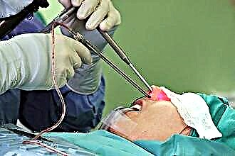
Conservative therapy consists in the use of broad-spectrum antibiotics, relief of the outflow of pus, parallel treatment of the nasopharynx and mucous membranes of the paranasal sinuses. If there are signs of a destructive stage, surgical intervention is immediately carried out. It consists in trepanning the mastoid process and removing all affected tissues through an incision behind the auricle. Endotracheal or local infiltration anesthesia is used. With a normal outcome of the operation, the wound heals in 3 weeks. However, surgery can sometimes damage the facial nerve, especially in children.
Glomus tumor
A glomus tumor of the middle ear is a benign neoplasm that is localized on the wall of the tympanic cavity or bulb of the jugular vein, and is formed from glomus bodies. It cannot be completely removed. Despite its benign nature, the tumor can grow and affect healthy tissues, including vital organs (brain stem, medulla oblongata, blood vessels), which can be fatal.
Signs of a glomus tumor are a pulsating red mass behind the eardrum, facial asymmetry, hearing impairment, and dysphonia.
For a more accurate determination of the localization and size of the formation, MRI, CT, angiography and histological examination are used.
Sometimes, at first, embolization (cessation of blood supply) of the neoplasm is carried out, which leads to a suspension of its growth.After that, the tumor is surgically removed (in whole or in part). A gamma knife or radiation therapy is also used. A positive result is more likely with early detection. Timely intervention can significantly improve the patient's quality of life.