For infectious diseases affecting the perichondrium, there is a general group name - "perichondritis", which is adjacent to the indication of the localization of inflammation: perichonditis of the auricle. The disease can lead to necrosis of the cartilage tissue and subsequent deformation of the ear. Inflammation is accompanied by skin redness, swelling, swelling, and soreness.
In the later stages, according to the characteristic manifestations, the disease is easily diagnosed. However, in the initial stages, it can be confused with either an othematoma (accumulation of blood in the perichondrium) - with serous chondroperichondritis of the auricle, or with erysipelas - with a purulent disease.
Causes and mechanism of development of the disease
 Among bacteria - causative agents of the disease - perichondritis is most often caused by Pseudomonas aeruginosa, less often by green streptococcus, Staphylococcus aureus and other species. As a result of infection, the perichondrium is damaged. Therefore, with self-diagnosis, one of the characteristic signs that pay attention to is an inflammatory process that spreads to all overgrown shells, but does not affect the lobe.
The perichondrium - perichondrium - is a dense connective tissue membrane that covers most of the cartilage (auricle, larynx, costal hyaline, etc.) and serves as their nutrition, thanks to the network of blood vessels enclosed in it. The lower layers of the perichondrium, using cellular elements, contribute to the transformation of cartilage into bone.
Infection in the perichondrium can enter in two ways:
- through any damage from the outside - the primary type,
- from internal infected organs with blood flow - secondary type.
Risk factors and preventive measures
Disease prevention correlates with avoiding major risk factors and conditions that can provoke pathological activation of pathogens. When a primary infection enters, risk factors include:
 pet scratches
- insect bites
- frostbite and burns,
- operations with violation of the sterility regime,
- cosmetic procedures,
- piercing.
 pet scratches
In this regard, the fastest and most complete antiseptic treatment of injuries and injuries, regardless of their degree, is considered preventive measures. Even a minor scratch requires the application of antiseptic rules. If, nevertheless, an infectious focus has arisen, it should be eliminated as quickly as possible.
A secondary infection can be triggered by a general decrease in immunity, as well as:
- diabetes,
- Chronical bronchitis,
- bronchial asthma,
- rheumatoid arthritis,
- any infectious processes and complications after diseases (otitis media, flu, tuberculosis).
In this case, prevention is reduced to strengthening the immune system, complete completion of treatment, and also - to conduct adequate therapy. So, for example, in the treatment of purulent otitis media, surgical intervention before the complete destruction of the Pseudomonas aeruginosa is considered undesirable.
Symptoms of perichondritis
 Depending on the form of perichonditis, the symptoms may be less pronounced (serous type), and pronounced against the background of the rapid course of the pathological process (purulent type).
The serous, more rare, form develops most often as a result of the penetration of a weakly virulent infection after an insect bite, scratch or burn. It manifests itself accompanied by the following symptoms:
- redness of the ear with a characteristic glossy glossy shine,
- the successive appearance of swelling, edema and swelling, which first increases, and then, when denser, slightly decreases in size,
- manifestation of painful sensations that are present, but are not very pronounced,
- an increase in the temperature of the skin, which rises at the site of inflammation.
A purulent, more common, form causes vivid manifestations in the form of:
- first - tuberosity and uneven swelling,
- then - the spread of edema to the entire area of the auricle, except for the lobe (while the tuberosity is smoothed out and becomes invisible),
- the occurrence of intense localized, and later - diffuse pain, which at the first stage increases with palpation, and at the second stage it spreads into the cervical, occipital and temporal regions.
At the same time, the color of the skin changes - from red to cyanotic, a feverish state with a temperature of up to 39 C occurs, sleep and appetite deteriorate, and irritability occurs.
A test action is a short sharp pressure on the ear, in which the infiltrate (cell clusters with inclusions of blood and lymph) begins to oscillate. This fluctuation indicates the accumulation of pus and the beginning of the process of purulent softening of the tissue, which at later stages leads to the detachment of the perichondrium and the melting of the cartilaginous framework.
 To improve the diagnosis and differentiation of hematoma from perichondritis, as well as the serous form from purulent, diaphanoscopy (transillumination) is performed. The essence of the method is to transilluminate tissues (cysts and peri-cutaneous formations) with a beam of light. Transparent liquid, when translucent in a dark room, will have a reddish tint, cloudy - it will not shine through. The disease is determined by the color reaction:
- a light yellow color is given by the serous form,
- blackout - purulent,
- red color is visible with hematoma.
Treating inflammation
Alternative methods of treatment of perichondritis of the auricle should be avoided, since with late diagnosis and untimely initiation of antibiotic therapy, an unfavorable prognosis describes irreversible deformation of the auricle. Medical therapy can be carried out in physiotherapeutic, drug and surgical formats.

Physiotherapy
Physiotherapeutic procedures (including at home) are carried out only with serous perichondritis and during periods of exacerbation attenuation. With a purulent form, they are contraindicated. In addition to laser therapy, ultraviolet radiation, microwave and UHF, X-ray therapy (less often), the patient is prescribed adequate nutrition and maximum rest. However, physiotherapy measures are prescribed only in addition to antibiotic therapy.
Drug treatment
 This treatment takes into account two factors:
- the need for both local and systemic antibiotic therapy,
- the choice of the drug should take into account the type of pathogen: tetracycline, oxytetracycline, streptomycin, erythromycin, etc. are used against Pseudomonas aeruginosa, since this bacterium is insensitive to penicillin.
Oral administration of antibiotics as part of general therapy is possible according to the following schemes:
- Levofloxacin 250 mg (1 / day) for 10 days + Azithromycin 500 mg (one hour before meals) for 5 days.
- Amoxicillin + clavulanate 625 mg (3 / day before meals).
- Erythromycin 250 thousand units per appointment (4-6 / day).
To relieve pain, you can take anti-inflammatory nonsteroidal drugs and analgesics.
Injection method schemes:
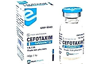 Cefotaxime 2 / day, 2 g, intravenously for 10 days.
- Streptomycin, 2 / day, 250 thousand intramuscularly.
 Cefotaxime 2 / day, 2 g, intravenously for 10 days.
In local antibacterial therapy, ointments with 2% mupirocin (10 days), 1% polymyxin M (5-10 days) are used. In addition, a 10% solution of silver nitrate (lapis) or a 5% tincture of iodine is used, but not together, since under the influence of iodine, lapis precipitates. Boric acid powder, which is blown into the ear canal, is especially effective against Pseudomonas aeruginosa, but boric acid can also be used in a solution in the form of a compress.
With the serous form, conservative methods are often sufficient. As a rule, on the third day there is a significant relief and improvement in the condition, however, it is important not to be deceived by this, to complete the course. In the case of a purulent form, surgical intervention can be dispensed with only in the early stages.
Surgery
 The basis for surgical intervention is fluctuation and aggravation of suppuration. To divert a small amount of pus, incisions with drainage are used in the places of disclosure. With significant inflammation, the following sequence of actions is performed:
- A wide incision is made parallel to the contour of the auricle to avoid deformation during subsequent scar formation.
- Pus, granulation and necrotic tissue are removed.
- Rubber drainage is laid.
- Three times a day, the wound is washed with antibiotics and antiseptics.
- Antiseptic dressings with ointment are changed several times a day (as needed).
- After the discharge has ceased, the drainage is removed, and a tight bandage and tamponade is applied to the ear to prevent narrowing of the ear canal.
- The patient is observed by a doctor until complete recovery.