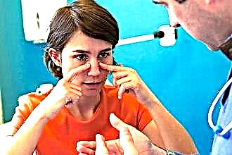
Neoplasms in the organ of hearing are most often discovered by a person unexpectedly, if they are not accompanied by painful sensations. This happens when cleaning the ear canal, washing or changing earrings. The appearance of a ball or lump in the earlobe, whether it hurts or not, does not always threaten health, but it can be a sign of atheroma, which can become inflamed.
Diagnosis of the disease
 If a ball appears on the earlobe or a lump has jumped up and hurts, then depending on the type of seal, the place of occurrence and specific features (mobility, soreness, change in the color and temperature of nearby skin areas), this may be:
- Traumatic swelling. If after the piercing pain and inflammation appeared, this means that an infection has joined the mechanical damage. The lump turns red and becomes painful, the temperature rises, and pus appears. Treatment with anti-inflammatory drugs is required. Also insect bites can cause such a tumor.
- Simple pimple or inflammatory infiltration. A lump on the tip or on the auricle is provoked by suppuration of the follicles of the hairs and skin glands. It can be a simple pimple or boil with a pronounced necrotic core.
- Epidermoid cyst. The result of abundant multiplication of epidermal cells, which collect in a seal, a ball in the earlobe, which only a physician can determine for sure. Able to fester, hurt and grow in size. It is similar in symptoms to the most common disease, which is described below.
- Atheroma (wen, sebaceous cyst). The most common disease, externally and to the touch, characterized by seals, bumps, balls in the earlobes. It is worth dwelling on it in more detail.
What is atheroma and the causes of its occurrence
Fat (atheroma) is a globular formation of a sebaceous gland that is clogged and stretched. This gives the impression that a ball has formed inside the earlobe. Mostly adults aged 25-50 suffer from atheroma, it is very rare in children and adolescents.

The sebaceous glands are microscopic in size, produce sebum, and are located across the entire surface of human skin. When the gland is blocked, it continues to produce a secret and, without proper treatment, can reach several centimeters in diameter. It usually looks like a hard ball (lump) in the earlobe, elastic, mobile and painless when not inflamed. The clots inside are a cyst filled with a yellow or white curd-like mass and lined with an epidermal layer. Clots can occur in an amount of 2-4 pieces, while the lymph nodes are often inflamed.
 A lump or wen in (on) the earlobe in the back or in the middle can occur due to one of the following reasons, although often even a specialist cannot say for sure:
- hereditary predisposition;
- hormonal disorders due to diseases of the endocrine system or age factors;
- changes in metabolic processes in the human body;
- acne;
- follicle injury, leading to complication of the outflow of secretion;
- seborrhea;
- excessive sweating;
- inaccurate ear piercing with subsequent non-compliance with hygiene rules;
- wearing jewelry made of low-quality materials that can react with liquids;
- prolonged stay in unfavorable conditions (for example, working in a hot, dusty room).
Symptoms of atheroma
 In fact, atheroma is a benign neoplasm. Education is small at first, but over time it can grow up to 5-6 centimeters and respond with pain. Clinical characteristics of common atheroma:
- well-defined boundaries;
- round form;
- smooth surface;
- mobility;
- the presence of an enlarged, edematous excretory duct in the center of the formation;
- filling the capsule with a mushy mass from the secretion of the gland and epithelial cells.
However, if a ball has formed on the earlobe, a large lump has jumped up and hurts, it means that the wen has become infected. The main features of this process are:
- soreness, especially when touched;
 hyperemia;
- high temperature;
- the outcome of the ichor with traces of pus and blood;
- the release of the contents of the capsule during its spontaneous opening.
 hyperemia;
Despite the fact that a neoplasm has appeared in the organ of hearing, people rarely go to the doctor, since they do not experience any unpleasant sensations and remember the cyst only when washing or cleaning the organ of hearing. However, at the initial stage, before the onset of the inflammatory process, the disease is much easier to treat.
In addition, an enlarged wen is a rather unaesthetic sight, especially for women. Without treatment, it can open up on its own over time and never bother again, but there is a danger of regular suppuration due to the remaining inside the capsule.
Traditional treatment of the disease
There is only one method of treating neoplasms of this kind (wen, epidermoid cysts) - surgical removal. The reasons for the intervention are prevention of suppuration and a cosmetic factor. Removal of a neoplasm is possible in several ways:
- Radio waves of high frequency, under the influence of which the contents of the adipose tissue and epithelial cells evaporate, while the surrounding tissues do not heat up. Local anesthesia is used, no stitches are needed.
- Laser moxibustion. Like radio waves, it can be used only in the early stages of the disease, when compaction is not more than 5 mm in diameter.
 Surgical removal with a traditional metal scalpel.
 Surgical removal with a traditional metal scalpel.
Operations of this type are nowadays carried out in polyclinics under local anesthesia. A thin incision is made with a scalpel and the contents of the cyst are extracted. In a successful case, the surgeon manages to remove the whole capsule, but if the capsule breaks, then it is necessary to remove it piece by piece. At the end of the manipulation, stitches are applied for several days. With timely intervention, there are practically no traces of the operation; a scar will remind of a delay in the trip to the hospital.
Sometimes, with severe suppuration, you have to resort to surgery twice. First, the wen is opened and the contents are removed, which is sent for histological examination to confirm the benign quality of the neoplasm. After removing the inflammation with medication, a second operation is performed, during which the capsule itself is removed. If this is not done, then relapses are possible.
In parallel with surgery, anti-inflammatory drugs are used, and when the temperature rises above 38 degrees, antipyretic drugs are prescribed.
Folk remedies
 Treatment with traditional medicine in case of infection of atheroma should not be abused in order to avoid the ingress of pus into the bloodstream during the natural opening of the seal. The most commonly recommended methods are:
- Aloe leaves are applied to the affected area 2 times a day for a month. They help to draw out the contents of the bud.
- Lamb fat is heated, cooled and rubbed in the right place.
- Garlic is chopped, mixed with sunflower oil. The product is rubbed in with light massage movements. If you feel a burning sensation, you need to repeat the procedure, reducing the amount of garlic.
- Salt, yogurt and honey are mixed in equal proportions and applied to the wen.