Diseases of the cardiovascular system in the 21st century occupy a leading position among the causes of death of the population. Left ventricular anterior wall infarction is the most common type of damage localization. It disables millions of people every year and, if not treated promptly, can be fatal. But is there any need to panic? We will discuss this issue below.
Causes, risk factors and development mechanism
To begin with, I will try to briefly outline the essence of the problem. Acute myocardial infarction is one of the variants of coronary heart disease (CHD). The basis of the pathology remains a critical discrepancy between the needs of the heart muscle for oxygen and nutrients. Simply put, individual parts of the myocardium are simply not supplied with blood, which leads to the death of first isolated cells (cardiomyocytes), and then entire areas of muscle tissue.
My patients often ask me what can happen to the vessel that feeds the heart with blood closes. In 70-80% of cases, the problem is atherosclerosis. The disease is accompanied by the deposition of fat on the surface of the vascular wall and inflammation of the cells of its inner layer. Over time, these elements form a tubercle, after which there is a narrowing of the lumen of the artery. The result can be their rupture of plaque with the formation of a blood clot and complete blockage of the vessel. Acute myocardial infarction of the anterior wall of the left ventricle occurs in my practice more often than other forms. This is due to the anatomical features of the blood supply to the heart.
According to the modern views of cardiologists on the causes of the development of myocardial infarction, it is worth highlighting the following key factors that contribute to the progression of pathology:
- Improper nutrition. I mean excessive consumption of foods rich in fast carbohydrates and fats (fast food, various sweets, energy drinks, alcohol).
- Obesity. Excess body weight indicates an active process of fat deposition in the vascular wall. You can determine your body mass index by following the link.
- Smoking. Nicotine contributes to additional spasm of the coronary arteries.
- Physical inactivity. The less you move, the higher the chance of developing obesity with further progression of atherosclerosis.
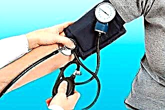
- High blood pressure (BP). The chance of developing anterior, lateral, or inferior myocardial infarction in hypertensive patients is higher than in patients with normal blood pressure.
- Stress. Emotional stress leads to vasospasm, which, against the background of the presence of plaques, increases the risk of rupture with the development of acute thrombosis and blockage of the artery.
- Age, gender. Men over 50 occupy a leading place among patients with heart attacks of different localization.
Genetic predisposition also plays a role in the development of the disease. If you have close relatives who have suffered or died from a heart attack, you should be more careful about risk factors and visit a doctor more often.
Symptoms
An infarction of the anterior wall of the heart (as well as other localization) is accompanied by the appearance of characteristic clinical signs that allow one to immediately suspect the disease.
Typical signs
When talking with patients suffering from hypertension or other heart diseases, I always pay attention to the signs that may indicate the onset of the development of acute infarction of the anterior wall of the left ventricle:
- Burning, pressing pain in the region of the heart. Many of my patients have described the symptom as "squeezing in a vice" or "an elephant stepped on the chest." A feature of a heart attack is the irradiation (spread) of pain to the left arm, neck, jaw or under the scapula.
- The duration of the attack may exceed 30 minutes.
- The ineffectiveness of the pills used. My patients always keep antihypertensive medicines, Nitroglycerin and Validol at home. In case of a heart attack, these drugs will not bring relief.
Most of my patients can clearly indicate the moment of pain. The attack is often associated with a stressful situation or excessive physical exertion. However, in my practice, there have also been patients who have developed a heart attack in the absence of any provoking factors.
Atypical manifestations
The clinical picture described above remains classic. It is typical mainly for anterior infarction. However, plaque rupture with thrombosis can also occur in arteries that supply blood to other parts of the heart.
With myocardial infarction of the lower wall of the left ventricle, the following atypical signs come to the fore:
- Nausea, vomiting. One patient told me how he ate a heavy meal and his abdominal discomfort began to grow sharply. Before seeking help, he drank 4 tablets of drugs to improve digestion. The ambulance team diagnosed inferior infarction.
- Isolated shortness of breath with a tendency to increase the intensity of the symptom. In this case, we are talking about the asthmatic "mask" of the disease.
- Weakness with episodes of dizziness. Patients rarely lose consciousness.
- Painless form of ischemia. A relatively rare variant of the development of the disease. The patient may report exclusively weakness and a desire to rest.
I always tell my patients that if at least one of the symptoms described above occurs, it is worth contacting a specialist. It is quite easy to skip a heart attack that develops in the lower wall of the left ventricle. However, treating it after losing precious time is not easy.
Diagnostics
Inferior, posterior, or anterior myocardial infarction is diagnosed in the same way. First, I always take a medical history and assess the patient's complaints. More often than not, chest pain alone is enough to raise suspicion.
To confirm the guess, I use auxiliary instrumental and laboratory examinations.
Instrumental methods
The basis for the diagnosis of any myocardial infarction is the ECG. It is impossible to overestimate the value of the electrocardiogram in IHD. The technique allows you to see the slightest deviations in the electrical function of the heart on paper or on a screen, which always occur when there is a violation of the supply of certain parts of the myocardium with blood.
Possible changes on the film:
- elevation (rise) or depression (subsidence) of the ST segment relative to the isoline;
- inversion (change of polarity to the opposite) of the T wave;
- the formation of a deep and wide (pathological) Q wave.
There are indirect signs on the ECG, which may indicate anterior infarction or damage to the other wall of the left ventricle.
To clarify the location and degree of damage to the heart muscle, I always additionally prescribe the following studies:
- Angiography of the coronary vessels. After the contrast is injected into the coronary arteries, I can see the blockage on the monitor screen, which makes it possible to quickly restore the vessel patency by stenting.
- Echocardiography (Echo-KG). Ultrasound examination of the heart allows you to see a decrease or complete absence of contractions of the affected area of the myocardium (hypo- or akinesia).
In 98% of cases, the instrumental techniques described above are sufficient to make the final diagnosis.
Laboratory methods
Laboratory tests are excellent helpers in the early stages of disease verification. The most reliable is a blood test for troponin I. The latter is a protein contained in cardiomyocytes. With the death of myocardial cells, troponin enters the bloodstream, where it can be fixed.For more information on how to do it, read the article at the link.
Additional laboratory tests:
- General blood analysis. With a heart attack, the number of leukocytes may increase, and the erythrocyte sedimentation rate (ESR) may increase.
- Blood chemistry. The amount of C-reactive peptide, AST, ALT may increase.
- Coagulogram. The analysis demonstrates the function of blood clotting. In heart attack patients, it is often too pronounced.
Among laboratory tests, I, like the majority of cardiologists, primarily do an analysis for troponin. Other tests are of a secondary nature.
Consequences and possible complications
The prognosis for a patient with a heart attack always depends on a timely visit to a doctor. With the provision of qualified assistance to the patient within the first 2 hours after the onset of the attack, it is likely that the development of necrosis of the heart muscle will be prevented. A similar prognosis is available thanks to rapid thrombolysis and stenting. However, people often endure pain, hope that "it will pass by itself," thereby losing precious minutes and increasing the area of the lesion.
The most common complications of the disease that I often meet:
- Deterioration of the contractile function of the heart with the development of failure.
- Various kinds of rhythm and conduction disturbances.
- Chronic aneurysm of the heart. Due to the thinning of the affected myocardium, a bulge forms in the wall, in which blood clots can form.
The most severe consequence of a heart attack is death. However, provided adequate therapy and a successful combination of circumstances, patients can live well for decades even after suffering a heart stroke. You can read about what medications and how long to take after discharge from the hospital here.
Expert advice
My advice to patients is quite simple:
- quit smoking;
- less nervous about trifles;
- rationalize food: you do not need to give up your favorite dishes, the main thing is moderation;
- regularly undergo preventive medical examinations;
- move more and engage in feasible physical education.
It is almost impossible to completely protect yourself from a heart attack. However, thanks to the basic points indicated above, it is possible not only to improve well-being, but also to prevent the progression of more than two dozen internal diseases.
Clinical case
A 49-year-old man was admitted to our clinic with severe pressing pain behind the sternum, which radiated to the left arm. The patient associates symptoms with stress due to an argument with his wife. From the moment the symptom appeared to seeking help, 2 hours passed. On the cardiogram, there is ST segment elevation in V1-V4 and the formation of a pathological Q wave in I, aVL, V1-V4. When conducting bedside Echo-KG zones of hypokinesia were not revealed. The troponin test is positive. BP - 130/90 mm Hg. Art.
The patient is referred for urgent coronary angiography. Total occlusion of the anterior descending branch of the left coronary artery was found. Stenting with a metal stent was performed. As a result, the diagnosis was made: antero-septal myocardial infarction. On the third day after stenting and taking appropriate drug therapy, the patient noted almost complete normalization of the condition.