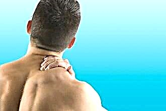
The functions of the larynx, which is a hollow organ connecting the hypopharynx (above) with the trachea (below), are not limited to providing air access to the lower respiratory tract. The structure of the human larynx allows one to speak with emotional shades, loudly and quietly, to sing, creating sounds of different heights and power. But the main task of this organ is to protect the lungs from the ingress of foreign bodies, for which nature provides for a reflex mechanism of laryngeal contractions and coughing.
Functions and biological capabilities
Functionally, the human larynx is an organ that solves several problems at once, and there are still different theories regarding the mechanisms for solving some of them.
There are four main functions of the larynx:
- Protective. Protection is provided by the movement of the cilia that cover the mucous membrane. Cilia capture even small dust particles trapped in the respiratory tract. Then the dust is surrounded by phlegm and when the reflex coughing mechanism starts, it is excreted from the body along with the mucus. This process takes place continuously.
- Respiratory. When inhaling through the mouth or nose, the air sequentially passes through the pharynx, larynx, trachea, bronchi, reaching the lungs, and when exhaling, it returns in the opposite direction.
- Voice-forming (phonatory). Oscillation of the vocal cords on exhalation causes a sound, the characteristics of which depend on the width of the glottis and the tension of the ligaments.
- Speech. This function is described only in conjunction with the work of the organs of the oral cavity (lips, tongue, teeth), while mentioning the functions of the chewing and facial muscles, which ultimately ensures articulate speech. Therefore, sometimes the speech function of the larynx is excluded from the list.
Protective function
The protective functions of the larynx are associated with the location in the organ of three reflex zones of the mucous membrane:
- The first zone surrounds the entrance to the larynx (the mucous membrane of the scapular folds, the surface of the epiglottis).
- The vocal fold is the location of the second zone.
- The location of the third zone is the subglottic cavity of the larynx on the cricoid cartilage.
The receptors of these zones are sensitive to temperature, tactile, and chemical influences.
When the mucous membrane of these areas is irritated, a spasm of the glottis occurs, which blocks the underlying airways and protects them from foreign objects, food, saliva. And with irritation of the reflexogenic areas and the sub-vocal space, a reflex cough occurs, with which foreign objects are pushed out.
At the entrance level, by analogy with the railway switch, there is a division into two directions: into the digestive and respiratory tracts. At the moment of swallowing, the larynx rises to the root of the tongue, moving up and forward, and the epiglottis bends towards the back of the pharynx so that it blocks the entrance to the larynx. Food from both sides flows around the epiglottis, falling into the pear-shaped sinuses, and then into the esophagus that opens at this moment. At the same time, during the act of swallowing, the arytenoid cartilages tilt and the vestibular folds close.
Respiratory function
Through the larynx, air passes into the lower trachea, bronchi, and lungs. When inhaling, the glottis expands by an amount depending on the needs of the body. The deeper the inhalation, the stronger, and with a deep inhalation, the bifurcation of the trachea is often visible. Such expansion is always a reflex process, which is due to the effect of the inhaled air on the nerve endings of the mucous membrane. The impulse from them through the afferent fibers of the superior laryngeal nerve and then through the vagus nerve passes to the respiratory center, which is located at the bottom of the fourth stomach. After that, the motor impulse enters the muscles that expand the glottis through the efferent fibers. At the same time, the functions of the muscles of the diaphragm and intercostal muscles participating in the act of breathing are activated.
Phonator function
The mechanics of sound reproduction involves the participation of all parts of the respiratory apparatus:
- lower resonator - lungs, bronchi, trachea,
- parts of the larynx in the part of the vocal apparatus,
- upper resonator - cavities of the paranasal sinuses and nose, pharynx, mouth (with the ability to change shape due to the movement of the lips, cheeks, lower jaw).
The anatomy of the larynx assumes the following process of sound formation: the closed glottis is opened by the pressure of air coming from the lower resonator, due to the elasticity and elasticity of the vocal folds, after which the return phase begins, during which the slit closes again. The folds vibrate perpendicular to the outgoing air stream. And the frequency of these vibrations corresponds to the pitch.
The pitch is determined by the number of vibrations of the vocal cords in one second.
- Breast register. To evoke sounds of a certain pitch, a person, with the help of a reflex mechanism, "sets" the necessary tension and length to the folds, as well as the corresponding shape for the upper resonators. The pattern of oscillatory movements of the vocal folds resembles the vibration of a steel ruler, which performs oscillatory movements and one end of which is fixed, and the other is free. The oscillation of the extended and released ends produces a sound, but in the case of a laryngeal extraction of a sound, the force is regulated arbitrarily.

- Falsetto. With falsetto, the glottis does not close completely, therefore, the air, with effort passing through it, vibrates the edges of the adjacent folds. In falsetto, the folds are flat, strongly stretched, but the sound is weaker than the chest one. ...
- Whisper. In this version, the folds are closed 2/3 in front, and a triangular slit remains in the back. The air passing through it causes a quiet noise - a whispering voice.

The width of the slit is regulated by at least five laryngeal muscles, expiratory force, and other factors. The fact that sound arises due to the "work" of the throat and larynx was known even in the time of Hippocrates, but only 20 centuries later, in the 16th century, Vesalius suggested that the vocal cords play the main role in this process. But the theory of sound production is being finalized today. Now they are talking about two theoretical options:
- Aerodynamic theory describes the process of voice formation as a result of vibrations of the vocal folds, where the muscles involved in the exhalation phase and the muscles of the larynx, which bring the ligaments closer together, play the main role. But these muscles are triggered reflexively at the moment of irritation of the mucous membrane with an air stream.
- In the second variant, the muscles are "switched on" not passively, but on a command from the brain, which came through the nervous system.
Interesting facts about the laryngeal "instrument"
In the course of development, the sex and age characteristics of the larynx are revealed. Up to 10 years old, boys and girls have almost no differences in its structure. In newborns, it is located three vertebrae higher than in adults, representing a wider (especially at the entrance) and short cavity. At an early age, a large amount of loose connective tissue is concentrated in the lining space, which provides an environment for the development of edema (lining laryngitis, false croup, etc.).
The cartilage of the larynx, which consists of hyaline cartilage (and this is all, except for the epiglottis) from 25-30 years old, soak in calcium salts. From this age, ossification progresses, and by about 60-65 years, ossification becomes complete.
The larynx, the anatomy of which is only formed by the age of 7 years, at a young age does not yet have cartilage cartilage and thyrohyoid ligaments.But before the onset of puberty, the production of male sex hormones in boys and the active development of the gonads leads to rapid growth and significant lengthening of the vocal cords (12-15 years). This is also associated with a voice mutation lasting about a year and ending by the age of 14-15. In girls, growth occurs gradually, and the voice "breaks" quickly and imperceptibly by the age of 13-14.
To preserve the pure high sounding of the boys, who were supposed to sing in the papal choir, in Italy of the 17th – 18th centuries. the castration procedure, performed at the age of 7-8 years, was widespread. Due to this, puberty had little effect on the size of the laryngeal cavity, which provided a combination of masculine strength, high tone and neutral (between masculine and child) timbre.
Normally, the larynx - structure, function, degree of development - retains gender differences even in adulthood: the male cavity is about a third larger than the female, the vocal cord is one centimeter longer and much thicker. This determines the properties of a stronger and lower male voice. On average, the vocal folds of newborn babies are 0.7 cm long, female - about 1.6-2 cm, male - 2-2.4 cm.The width of the glottis in the posterior third between the vocal folds is on average 15-22 in men. mm, in 10-year-old children - 8-11 mm, in women - 13-18 mm.
But, in this case, a “strong” male voice can only be called by comparing it with a female or a child's, since the vocal sound energy is so small that if a person speaks continuously for 100 years, then the heat energy will only be enough to cook one cup of coffee.
Features of the anatomical structure
Anatomically, the larynx is a complex complex of anatomical and physiological elements and tissue structures, served by a developed system of blood vessels, lymph vessels, and nerves. From above, the organ is attached to the hyoid bone by the thyroid-hyoid membrane, and below it is connected to the trachea with the help of the signetracheal ligament. The upper edge is located at the level between the IV and V cervical vertebrae, the lower - at the level of the VI-th. Behind, the larynx is bordered by the entrance to the esophagus and the laryngeal part of the pharynx.
Active up and down movements, which are performed by the organ during breathing, swallowing, speaking, singing, are complemented by passive displacements to the right and left with crepitus (crisp sound) of the cartilage of the larynx. With a malignant tumor lesion, active and passive mobility is markedly reduced.
In the upper section of the thyroid cartilage in men, the Adam's apple (Adam's apple) is felt and visualized, which is less pronounced in women and children and has a soft formation in them, which greatly complicates its palpation.
At the same time, in all people, the area of the conical ligament is easily felt in the front in the lower section between the cricoid and thyroid cartilages. It is she who is dissected in case of asphyxia with an urgent need to restore breathing.
Outside, the larynx is covered with skin, subcutaneous tissue, superficial fascia (connective membrane) of the neck and muscles:
- the lateral parts of the fascia of the thyroid gland, which is attached to the lower part of the cricoid cartilage, are protected by the muscles of m. sternothyroideus et m. sternohyoideus,
- the anterolateral surface is covered by the sternohyoid muscle, with the sternohyoid and thyroid hyoid located under it.
Internal structure
The structural features - the anatomy and physiology of the larynx - are described by the complex interaction of cartilage, muscles, joints, ligaments, nervous, blood, lymphatic systems.
The inner surface is covered with a thin mucous membrane, which consists of stratified epithelium. The larynx cavity is similar in shape to an hourglass, that is, it is narrowed in the middle section and expanded from below and from above. Therefore, the anatomy of the larynx is presented as a "three-story" formation, in which:
- the upper floor - the vestibule of the larynx - is located between the entrance and the vestibular folds (vestibular fold) and looks like a cone-shaped cavity with a narrowing at the bottom,
- the middle floor - the glottis - is located between the vocal folds,
- the lower floor - the sub-vocal cavity - extends to the trachea and looks like a cone-shaped cavity with an expansion below.
In front, the entrance is bounded by the epiglottis, from the sides - by the scooplary laryngeal folds, in the lower region of which there are wedge-shaped and rod-shaped cartilages, and in the back - by the tops of scooped cartilages.
Pear-shaped pockets are located between the folds and walls, passing into the esophagus. The lingual-supraglottic grooves (valecules) are located between the lateral lingual-supraglottic and median folds. On each side of the two horizontal pairs of folds (vestibule and vocal folds), located in the middle and lower third of the thyroid cartilage, there are depressions - the morganic ventricles of the larynx. Two upward pockets extend from them. The accumulation of lymphadenoid tissue in the thickness of the mucous membrane of the laryngeal ventricles is sometimes called the laryngeal tonsils.
The vocal fold passes into the elastic cone of the larynx, more precisely - it is the upper-posterior bundle of the elastic cone. It covers the vocal muscle stretched between the inner vocal process and the angular surface of the thyroid cartilage.
Laryngeal cartilage
- The device of the thyroid cartilage located on the cricoid cartilage is described as a 38 ° connection of the plates that protect the organ from external mechanical influences. On the corner at the top edge is the top notch. Paired thyroid-hyoid muscles (working on the rise) and sterno-thyroid (working on lowering) muscles are attached to the surface of the plates from the outside. The posterior edges of the plates pass into the lower and upper horns.
- The cricoid cartilage works as the base of the larynx. From below it is associated with the trachea, and from above - with the thyroid cartilage.
- Arytenoid cartilage, which is named after the configuration of the rowing movement of oars, has the shape of a triangular pyramid, which is located at the upper-posterior border of the cricoid cartilage plate. Each of these arytenoid cartilages has a vocal process with a vocal fold attached to it.
Laryngeal muscles
All laryngeal muscles are divided into external and internal. The internal muscles of the larynx, in turn, are divided into 3 groups:
- Muscles that dilate the glottis. It is represented by the only pair of posterior cricoid muscle innervated by recurrent nerves.
- Muscles narrowing the glottis (adductors). The group is represented by two paired (cricothyroid and thyroid) and unpaired transverse arytenoid muscles.
- Muscles that serve to tighten the vocal folds. The group includes paired thyroid and cricothyroid muscles.
The external muscles include three pairs:
- sterno-thyroid,
- thyroid-sublingual,
- lower pharyngeal compressors.
With the help of these muscles, the laryngeal position relative to the pharynx is regulated: during swallowing, the larynx rises, during breathing and sound extraction, it descends.
Ligaments and joints
The main ligaments of the larynx:
- Lateral and thyroid-hyoid median. It is a part of the thyroid-hyoid membrane, through the opening in which the neurovascular bundle enters. The median thyroid hypoglossal connects the body of the hyoid bone to the upper edge of the thyroid cartilage.
- Shield-laryngeal. Connects the thyroid cartilage in the upper border zone with the epiglottis.
- Sublingual epiglottis. Attaches the epiglottis to the body of the hyoid bone.
- Cricotracheal. Bonds the larynx to the trachea.
- Cricothyroid. As a continuation of the elastic membrane of the larynx, it connects the lower border of the thyroid cartilage with the upper edge of the cricoid cartilage arch.
- Cherpalonadlaryngeal. Located on the border of the lateral edge of the epiglottis and the inner edge of the arytenoid cartilage.
- Lingual-epiglottis median, as well as lateral.Tie the lateral and median parts of the tongue root on one side and the anterior surface of the epiglottis on the other.
Blood supply and innervation
The neurovascular bundles are located on the sides of the larynx. The blood supply and innervation of the larynx is carried out by two arteries and two branches of the vagus nerve.
Arteries that supply blood to the larynx:
- The superior laryngeal artery is a branch of the superior thyroid artery, which is a branch of the external carotid artery. The superior laryngeal artery is larger than the lower one. It supplies the organ as part of one of the neurovascular bundles through the opening of the thyroid-hyoid membrane. Further, there is a division into smaller branches, including a branch of the middle laryngeal artery, which connects to the artery of the same name from the opposite side, located in front of the conical ligament.
- The inferior laryngeal is a branch of the inferior thyroid artery starting from the thyroid trunk.
Through the superior thyroid vein with the transition to the internal jugular and then to the lower thyroid and brachiocephalic veins, venous outflow is carried out.
 The laryngeal lymphatic system is also divided by the vocal folds into the upper and lower sections. Moreover, the upper network is more developed (especially in the areas of the laryngeal ventricles and vestibular folds). From here, the lymph enters the deep cervical lymph nodes, moving along the line of the neurovascular bundle. The lymph nodes of the lower section are located above and below the cricoid cartilage, then grouping into the pre-epiglottis lymph nodes. The connection between the lymphatic system of the lower section and mediastinal lymph nodes is clinically significant.
In general, the lymphatic system is more developed here than in other organs of the neck (the ventricle and folds of the vestibule of the larynx are especially prominent). The least developed lymphatic system is in the area of the vocal folds, which causes a relatively late metastasis of cancer cells here.
The innervation of the muscles is provided by two branches of the vagus nerve:
- The upper one, which departs from the wandering in the region of the lower part of the node. On the back of the large horn of the hyoid bone, it divides into two more branches:
- the outer innervates the cricothyroid muscle,
- the inner one spreads sensitive branches to the mucous membrane.
- The lower one innervates all the internal laryngeal muscles, except for the cricothyroid, providing sensitivity to the lower mucosa along with the area of the vocal folds. The inferior nerves are an extension of the left and right recurrent nerves extending from the vagus at different levels in the chest cavity:
- right - at the level of the subclavian artery,
- left - in the area of the aortic arch by the vagus nerve.
Possible diseases
The specialist examines the condition of the laryngeal cavity using a laryngoscope, one of the main elements of which is a small mirror. The idea of this device in 1854 brought the singer and vocal teacher M. Garcia the title of honorary doctor of medicine. And although the diagnostic capabilities of medicine have expanded since then, doctors still use a laryngoscope for examinations. However, quite often the epiglottis, presented in the form of a half-rolled sheet in children of some adults, covers the laryngeal entrance and interferes with medical examination using the method of indirect laryngoscopy.
The main acute diseases of the larynx include:
- Acute catarrhal laryngitis is an inflammation of the mucous membrane. As an independent disease, it arises as a result of the activation of the flora under the influence of endogenous and exogenous factors.
- Infiltrative laryngitis is an acute disease associated with a bacterial infection, in which the inflammatory process is not limited to the mucous membrane, but spreads to nearby tissues with the involvement of the muscular apparatus, ligaments, perichondrium.
 False croup - acute laryngitis, characterized by the localization of the process mainly in the subglottic space. It is fixed in children under 6-8 years old, which is due to the specifics of the structure of the larynx, namely the sub-vocal space in the child. Developed loose fiber reacts quickly to infectious irritation. In addition, the disease is facilitated by the narrowness of the children's larynx, as well as the lability of nerve reflexes. With blood flow in a horizontal position, the edema increases, so the patient's condition worsens at night.
 False croup - acute laryngitis, characterized by the localization of the process mainly in the subglottic space. It is fixed in children under 6-8 years old, which is due to the specifics of the structure of the larynx, namely the sub-vocal space in the child. Developed loose fiber reacts quickly to infectious irritation. In addition, the disease is facilitated by the narrowness of the children's larynx, as well as the lability of nerve reflexes. With blood flow in a horizontal position, the edema increases, so the patient's condition worsens at night.
Infectious (with bacterial, fungal and viral pathogens) laryngeal angina affects the lymphadenoid tissue in the morganic ventricles, the surface area of the epiglottis, the area of the piriform sinus floor, etc.
The occurrence of certain physiological conditions can be judged by the changes in the voice.
In women, for example, voice changes can illustrate a change in hormonal levels during menstruation, menopause, and changes associated with taking hormonal drugs.