Identifying ear cholesteatoma can be challenging, even for a specialist, since the disease is asymptomatic in its early stages. However, complaints of the appearance of dull, aching, pressing or shooting ear pain, hearing impairment, and in some cases dizziness and the appearance of fetid discharge indicate the development of purulent-inflammatory diseases in the patient, which, in turn, often cause the formation of cholesteatoma.
Causes of occurrence
 Despite the fact that this pathology is similar to an ear tumor, their similarity is purely formal. Unlike a tumor-like formation, a cholesteatoma consists of several layers contained in a capsule of connective tissue. On the basis of its appearance - a smooth surface - it got its name "pearl tumor". Inside the shell there is a keratinized epithelium, cholesterol crystals and keratin, and the nucleus of this neoplasm is a white substance with a pungent unpleasant odor.
The nature of the occurrence of middle ear cholesteatoma is different. So, it can form as a result of trauma or neglected purulent diseases of the auditory organ - in 90% of cases, its occurrence is the result of chronic purulent otitis media. Cholesteatoma acquired during life is also called "false".
There are two types of mechanism for its appearance. In the first case, there is an ingrowth of the squamous epithelium of the outer ear into the middle cavity through a rupture in the tympanic membrane. In the second, a decrease in pressure in the tympanic cavity, provoked by Eustachitis, leads to the retraction of a rather large portion of the tympanic membrane into it, where keratin and epithelial particles begin to collect.
In rare cases, this disease can be congenital in nature, then it is called "true".
As a rule, embryonic disorders become the cause of congenital pathology, and it is localized in the pyramid of the temporal bone. In any case, this pathology entails serious damage to the middle ear, since as it grows, it begins to put pressure on the surrounding bone tissues, which provokes their destruction. In addition, cholesteatoma secretions are toxic and can lead to disturbances in sound perception. Thus, this disease can lead to serious complications in the form of brain abscess, meningitis, meningoencephalitis, paralysis of the facial nerves, etc.
Diagnostics
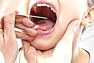
Timely diagnosis of this disease is an important factor for further effective treatment and prevention of complications. When the above-described clinical symptoms and signs appear (ear pain of a different nature, detritus discharge, headaches, dizziness, hearing impairment, etc.), otolaryngologists, as well as neurologists and neurosurgeons, resort to instrumental diagnostic methods. The most effective diagnostic procedures are:
- otomicroscopy;
- temporal bone radiography;
- CT scan;
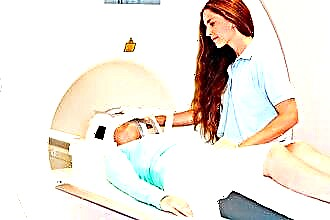
- Magnetic resonance imaging;
- audiometry (detection of hearing loss);
- tonal threshold audiometry (detection of mixed type of hearing loss);
- vestibulometry (analysis of the functions of the vestibular apparatus).
Treatment
In the early stages of the development of this disease, it is possible to use drug treatment. The basic principles of such therapy are the washing of the tympanic space with a solution of boric acid or proteolytic enzymes. If the usual method of washing does not help to improve the patient's condition, then a special drum-cavity tube with a bend at the end is used for this procedure, which is inserted through an opening in the tympanic membrane. With successful treatment, the patient has a cessation of suppuration, scarring, as well as regeneration of the tissues of the tympanic space.
However, in most cases, conservative treatment does not bring the desired effect, so the problem is solved mainly by surgical intervention.
The operation to remove the ear cholesteatoma is divided into several stages:
- direct removal of the "pearl tumor";
- sanitation of the cleaned middle ear cavity (in order to prevent the recurrence of the disease);
- restoration of injured auditory ossicles;
- restoration of the integrity of the tympanic membrane.
Postoperative period
Immediately after surgery, the patient may experience dizziness and nausea, but over the next 7-10 days, these postoperative symptoms gradually disappear. Before the patient is discharged from the hospital, the stitches are removed from the wound behind the outer ear. Then a dressing is made at this place, which must be changed every few days and completely removed after the wound has healed. Four weeks after the operation to remove the cholesteatoma of the ear, specialists conduct a control examination of the patient's hearing. If additional surgical intervention is needed to improve it, then it can take place no earlier than 6 months after the first.
With a successful completion of the treatment, it should be remembered that the operated auditory organ has increased sensitivity and must be protected from hypothermia and various infections entering it.