If in the parotid area (more often - behind the ear) a pinkish-gray rounded mass 0.5-50 mm in size has arisen and become inflamed, then it can be assumed that the gland of the lymphatic system has become inflamed, or, as people often say, the lymph node has become inflamed and enlarged behind ear.
A significant difference in the size, shape, localization, causes of enlargement and inflammation of the lymph nodes on the face near (near) the ear, raises a number of questions related to people's attempts to conduct an independent diagnosis. To assess the degree of risk to health and relate the altered state of the lymphatic system to possible immune, infectious, tumor pathologies, one must understand what a lymph node is, exactly where it is located behind the ear, and know the location of the glands in the parotid zone.
Lymphatic system function
 The lymphatic system is considered as a part of the immune system, which transports immune cells and regulates the elimination of toxins from the lesion through the lymphatic system. The lymphatic system includes ducts, trunks, vessels, capillaries, nodes through which infection spreads in the event of a primary lesion.
Inflammation of the lymph nodes behind or around the ears (when the glands of the parotid lymph group are inflamed and enlarged), most likely, indicates a lesion of an organ close to the group.
In this case, lymphadenitis is secondary in nature and is a consequence of the spread of the primary lesion. Less often, primary lymphadenitis is recorded, arising, for example, due to a violation of the integrity of the skin and the ingress of infection directly into the lymphatic system.
Lymph nodes are the peripheral organs of such a system. They function as a biofilter, representing rounded or oblong (sometimes ribbon-like) pinkish-gray formations. They are located in clusters (in groups of up to 10 pieces) along the lymph vessels and most often near large veins. Their surface is covered by a connective tissue capsule, and the supporting structures of the trabecula (beams) extend from it. The structural basis of education is the stroma, which includes:
- reticular connective tissue,
- fibers that form a three-dimensional network,
- several types of macrophages (phagocytic cells).
The cortical substance is located near the capsule, and the inner part consists of the medulla. In the area of the superficial cortex are follicles - lymphatic nodules. Lymph slowly seeps through the internal spaces (sinuses), as a result of which it is cleansed, and the lymph itself is enriched with antibodies.
 Foreign antigens brought by lymph provoke an immune response and an increase in lymphoid accumulations. Inside the formation, lymphocytes also mature, participating in the fight against foreign substances.
In the body of an adult, there are about six hundred nodes, of which only the submandibular, inguinal and axillary are normally probed.
Therefore, the fact that an enlarged lymph node behind the ear in an adult in itself speaks of pathology, and an enlarged gland in children causes less concern, since the formation of immunity in a child is a more active process, and the reaction of the cervical nodes is a common phenomenon, which after some that time will most likely pass without a trace.
The location and functioning of the lymphoid glands in the parotid region
Since the nodes are located in groups, a picture is most often observed when the lymph nodes on and behind the ear (ears) have become inflamed along the path of the spread of infection or cancer. The regional cervical lymphatic system, which includes the cervical, parotid, occipital and supraclavicular nodules, provide protection for the organs and anatomical elements of the head.
Around the auricle and in relative proximity to it, there are the following nodal lymph nodes:
- parotid (under the lobe),
- preauricular anterior (in front of the tragus),
- posterior (behind the shell and closer to it than the occipital).
In a condition characterized by an increase in nodular formations, an intermediate diagnosis of "lymphadenopathy" is made, which, after clarification of the reasons for the increase (inflammation), is specified. In the presence of localized lymphadenopathy, anatomical areas are examined, from which lymph flows into this nodal group. And at the same time, an examination of non-contiguous nodal groups is carried out in order to exclude generalized lymphadenopathy. In this case, the state is assessed by a combination of five main criteria:
- Soreness. The pain is caused by a rapid stretching of the formation capsule and an increase in its volume, as well as by an inflammatory process with suppuration and hemorrhage into the necrotic center (in case of a malignant lesion). However, the presence of pain in this case does not allow a diagnostic separation between malignant and benign diseases.
- The size. Most often, the size of the formation normally does not exceed 1 cm.And although this indicator does not make it possible to clearly diagnose the disease, the suspicion of a malignant tumor arises when the node is enlarged by more than 1x1 cm.
- Consistency. Cancer tumors are characterized by very hard, like a stone, seals. A softer consistency is the result of an inflammatory process or infection. Viral diseases are characterized by multiple small ("buckshot") nodules under the skin.
- Interconnectedness (conglomerate). A conglomerate is a group of nodules that, in response to an infectious or tumor pathology, demonstrates the connection between the elements of the conglomerate. Such conglomerates are characteristic of tuberculosis, lymphogranuloma venereum, sarcoidosis, and also for malignant diseases.
- Localization. The anatomical position of the inflamed elements of the lymphatic system in the perioustal region allows, first of all, to assume:
- local infections (including furuncle, carbuncle),
- rubella,
- pharyngitis,
- lymphoma,
- tuberculosis.
All these diseases manifest themselves in one form or another of lymphadenitis.
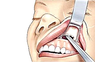
What if you find lymphadenitis in yourself? This is a serious symptom that cannot be ignored. It is very important to find out the factors provoking its occurrence. As soon as the cause is eliminated, the lump will instantly pass. Read more about what is the primary source of inflammation here.
Lymphadenitis manifestations
- Rubella. Rubella infection is regularly accompanied by the occurrence of lymphadenitis in the parotid, posterior cervical, occipital regions. Such inflammation is accompanied by a moderate increase, slight soreness, multiple or isolated asymmetric manifestations.
- Lymphomas. Hodgkin's lymphoma is characterized by lymphogenous metastasis from the primary focus to other nodal groups. The formations have a dense and elastic consistency, are painless and are not adhered to the skin. Non-Hodginsky lymphomas are also not adhered to the skin, dense and painless, but with them peripheral formations of the lymphatic system are much more often involved in the pathological process.
- Cat scratch disease. Taking into account the specific affected area, cat scratching affects, first of all, the cervical, parotid, elbow and axillary zones. The lesions are not adhered to the skin, however, they are painful and reach large sizes in diameter (3-5 cm).
Patient memo
- Is it possible to warm the lymph nodes behind the ear? In the event of an inflammatory process, heating will help to activate it, therefore, without a clear definition of the nature of the pathology, it is strictly forbidden to heat the nodes.
- Soldered lymph nodes behind the ear indicate the manifestation of acute lymphadenitis in a purulent form, in which (in contrast to the serous-purulent form) there is a gradual soldering (fusion) of the node with the underlying tissues. A similar picture of soldering can be observed in chronic abscessing lymphadenitis, which develops against the background of the previous productive form, in which soldering with the underlying tissues is not observed. In parallel with this, the density and soreness of the node increases.
- If, despite treatment with antibiotics and anti-inflammatory drugs, the nodes do not decrease, you should definitely consult a doctor to rule out lymphoproliferative disease. Particular concern should be shown with bilateral inflammatory processes in the behind-the-ear region.
- A decrease in the body's immune status, which occurs with avitaminosis, the use of immunocorrectors, impaired heat transfer, chronic diseases, become additional risk factors that contribute to the inflammation of the lymph nodes in front of, under and behind the ear or ears.
- After piercing (puncture in any part of the auricle), carried out in violation of hygienic rules, inflammation and enlargement of the lymph nodes on one side of the right or left ear also often begin - and more often the node under the ear (under the lobe) is inflamed.
 If a hard, inflamed lymph node behind the ear is swollen but elastic, it may indicate the presence of lymphoma.
- Inflammation of the lymph node (s) in front of the ear, when the preauricular node near the tragus has become inflamed, may occur due to micro-damage to the skin in the temporal part of the glasses arches. More dangerous and inflammatory scratches can be caused by pets (usually cats). However, most often in this zone, the lymphatic system reacts to the spread of infection due to the spread of a necrotic focus with furunculosis or complications of the carbuncle (serous-purulent lymphadenitis).
 If a hard, inflamed lymph node behind the ear is swollen but elastic, it may indicate the presence of lymphoma.