In otolaryngology, throat diseases are considered the most commonly diagnosed pathologies. This applies to childhood and adult patients. Most cases of acute diseases are recorded in the winter season, however, in some cases, the process becomes chronic, and therefore the symptoms are almost constant.
 Tonsillitis will be called chronic if there is an inflammatory process in the tonsils, and symptoms appear from time to time. Under the influence of some provoking factors, an exacerbation of pathology occurs, which is accompanied by an increase in the intensity of symptoms.
The palatine and other tonsils perform a protective function, as they are lymphoid structures and part of the immune system. Frequent attacks of pathogenic microorganisms against a background of weakened immunity lead to the development of acute tonsillitis, followed by a transition to a chronic form.
The risk of developing a chronic type of disease increases with a decrease in the body's resistance after suffering infectious diseases (measles, flu, scarlet fever), with hypothermia or exacerbation of chronic diseases.
Especially often the chronic form is recorded when the infection spreads from foci in the nasopharynx (sinusitis) or the oral cavity (caries).
In addition, chronicity is observed with improper treatment of the acute process, when inadequate antibiotic therapy is prescribed.
Antibiotic prescribing should be done exclusively by a physician based on the results of the antibiogram.
Of the predisposing factors, it should be noted:
- violation of nasal breathing with polyposis, adenoids, structural anomalies and curvature of the septum;
- insufficient oral hygiene;
- incorrectly selected bracket systems.
ENT throat diseases involving the tonsils are classified into:
- a simple form in which symptoms of a local nature are observed in the form of swelling of the mucous membrane, thickening of the arches, purulent masses and plugs appear in the gaps. Also, palpation of regional lymph nodes reveals lymphadenitis (enlarged, swollen, painful lymph nodes);
- toxic-allergic stage 1, when, in addition to local manifestations, rapid fatigue, malaise, periodic low-grade fever and dizziness are noted. Sometimes a person may notice arthralgia and cardiac pain, which indicates an exacerbation of the infectious and inflammatory process. It is worth noting that clinical signs from the heart do not cause changes in the electrocardiogram;
- toxic-allergic stage 2, at which changes in the ECG are recorded, a violation of the cardiac rhythm and constant subfebrile hyperthermia are detected. In addition, the pathology is characterized by functional disorders of the osteoarticular apparatus, kidneys and liver. During this period, the risk of exacerbation of pharyngitis, the occurrence of paratonsillar abscess, the formation of cardiac defects, the development of other infectious diseases, rheumatism and septic conditions increases. Sepsis is caused by the migration of pathogenic microorganisms along the bloodstream, which predisposes the emergence of distant infectious foci.

Symptomatically, chronic diseases do not manifest themselves with pronounced symptoms. A person may be bothered by tickling, scratching in the throat, the presence of a lump in the oropharynx, dryness and an unpleasant odor. After each exacerbation of tonsillitis, the improvement of the condition occurs extremely slowly, accompanied by the preservation of subfebrile condition and malaise.
Exacerbations in a simple form are recorded up to three times a year, and in the case of a toxic-allergic one - much more often, predisposing the formation of a paratonsillar abscess and the spread of inflammation to adjacent healthy tissues (laryngitis). The patient complains of subfebrile hyperthermia and constant weakness.
With chronic disease of the throat, the tonsils become a focus of infection, from which microbes spread throughout the body. Therefore:
- reduced immune defense;
- collagenoses are noted (dermatomyositis, periarteritis, lupus, scleroderma);
- skin diseases develop (eczema, dermatitis, psoriasis);
- nerve endings are affected (sciatica);
- autoimmune processes develop (vasculitis, thrombocytopenic purpura);
Diagnosis of a throat disease includes the collection of anamnestic information (frequent sore throats), examination by an otolaryngologist and additional studies.
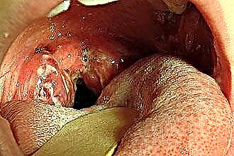
With pharyngoscopy, when the throat is examined, the disease manifests itself as reddening of the mucous membrane, thickening, as well as swelling of the arches. In children, loosening of the tissue of the glands is often found. Purulent discharge with an unpleasant odor accumulates in the lacunae. Palpation of regional lymph nodes reveals lymphadenitis (enlargement, edema, soreness of the lymph nodes).
Pharyngitis
Among the pathologies of the upper respiratory tract, pharyngitis is considered the most common. Previously, the acute process ended in recovery and did not lead to complications. Nowadays, patients with chronic diseases are increasingly turning to an otolaryngologist. In this case, the larynx is exposed to prolonged exposure to an infectious pathogen, which predisposes the persistence of inflammation.
When a sore throat is suspected, the chronic stage can take several forms:
- catarrhal, which is not characterized by a pronounced clinical picture;
- hypertrophic, which is characterized by the proliferation of mucous membranes and ridges;
- atrophic, when atrophy of the mucous membrane of the posterior pharyngeal wall occurs, which significantly disrupts its functions.
Sometimes there is a mixed form, in which some parts of the larynx are hypertrophied, while others have undergone atrophy, which is manifested by indistinct symptoms.
The reasons that provoke the chronization of the pathological process include viral pathogens (parainfluenza, adenoviruses, rhinoviruses) that persist for a long time in the mucous membrane. With frequent infection, the immune defense is so reduced that it cannot resist infection with streptococci or staphylococci.
As a result, even the slightest hypothermia or cold drinks can provoke an exacerbation of the disease. The disease becomes chronic against the background:
- smoking;
- alcoholism;
- inhalation of polluted air (smog, occupational hazards);
- immunodeficiency in severe chronic pathology;
- adenoids in children;
 closely located infectious foci (caries, sinusitis).
 closely located infectious foci (caries, sinusitis).
It is worth noting that chronic inflammation is possible with gastroesophageal reflux disease, when food is thrown, irritating the mucous membrane.
Allocate a sequential change in the mucous membrane, ranging from catarrhal symptoms, ending with atrophy.
Catarrhal form is often observed in smokers, as well as with the negative action of occupational factors. With the multiplication of microbes and the release of toxic substances, the appearance of puffiness and redness of the tissues is observed. Plaque from dead cells and pathogenic microorganisms can form on the surface.
In the future, there are large accumulations of mucus on the mucous membrane, which is difficult to cough up. The granular stage is considered the most dangerous in terms of the spread of infection.
| Stages | Symptoms | Picture with pharyngoscopy |
|---|---|---|
| Catarrhal | Discomfort, dryness, tickling, oropharyngeal tickling, soreness when swallowing, feeling of a lump | Not pronounced hyperemia of the posterior pharyngeal wall, slight thickening, swelling of the mucous membrane, the presence of thick mucus, which becomes more liquid during exacerbation. In some cases, the uvula and arches acquire an edematous and hyperemic appearance. |
| Granular | Vomiting, burning, severe cough. | Red nodules (granules) are visualized on the walls, plugs appear in the tonsils, the trigeminal nerve is irritated by granulosa clusters, and follicles are enlarged. |
| Hypertrophic | Discomfort, feeling of a lump, difficulty swallowing, drainage of mucus from the nasopharynx, frequent dry cough, unpleasant odor. | On the edematous, hyperemic walls, purulent mucus is located, the walls of the larynx and the lateral ridges of the arch are compacted. Purulent discharge can form crusts, and the granules gradually increase, provoking a transition to the atrophic stage. |
| Atrophic | Dryness, perspiration in the oropharynx, feeling of a lump, presence of crusts, cough, soreness when swallowing. | Sclerotic changes in the mucosa, as well as the submucosa of the pharyngeal wall and lymphoid structures. Thick mucus with a purulent component accumulates, forming dense crusts. The walls become thinned, pale, lacquered, through which fragile vessels are visualized. |
At the hypertrophic stage, tissue compaction is observed. With mucosal atrophy, crusts form, which can be released when coughing. Also, enlarged and painful lymph nodes on palpation are detected.
The disease in children passes in a catarrhal form, without causing clinical symptoms without exacerbation.
In adults, the disease manifests itself:
- unexpressed perspiration;
- small viscous secretions;
- the presence of a lump in the oropharynx;
- nausea, gagging when coughing up;
- dryness, irritation of the mucous membrane when inhaling cold air;
- rare cough;
- regional lymphadenitis;
- increased manifestations in the morning.
Complications are represented by the spread of inflammation to surrounding organs with the development of tracheitis, bronchitis or otitis media. In adults, there is a risk of transition of the catarrhal form to the hypertrophic and atrophic, disrupting the functions of the pharynx. Concomitant damage to the Eustachian tube leads to a decrease in auditory function.
In the diagnosis, analysis of anamnestic information, pharyngoscopy, laryngoscopy and laboratory tests (blood tests, smears) are used.
Pharyngomycosis
 The formation of an inflammatory focus caused by a fungal infection is called pharyngomycosis. Recently, otolaryngologists have noted the growth of fungal infection of the oropharynx. In most cases, pharyngomycosis is combined with stomatitis, gingivitis, or cheilitis.
It should be noted that fungal infection proceeds much more severely than bacterial inflammation and is less responsive to therapy. The cause of the development of the disease can be:
- yeast-like candida fungi, which cause thrush, candidiasis of the skin and genitals;
- molds (5%).
Activation and reproduction of fungal infection is noted against the background of immunodeficiency in HIV, frequent colds, tuberculosis or severe concomitant pathology (hypothyroidism, diabetes). In addition, the wrong course of antibiotic therapy, which exceeds the recommended dosages and duration, should be attributed to the predisposing factors. Also, pharyngomycosis is promoted by prolonged use of hormonal, chemotherapy drugs and removable dentures.
There are several forms of the disease:
- pseudomembranous, with a bark on the surface of the oropharynx, a white bloom is noted;
- erythematous, characterized by hyperemic areas with a smooth, varnished mucosal surface;
- hyperplastic - manifested by the formation of whitish plaques, which are difficult to separate from the mucous membrane, leaving a bleeding wound;
- erosive-ulcerative, when ulceration affects only the surface layers.
Sympathetically, the disease manifests itself as uncomfortable sensations in the form of perspiration, burning, dryness and tickling in the oropharynx. Soreness is not very pronounced, it increases with food intake, especially pickles and spices.
Painful sensations can spread to the ear and neck area. Lymphadenitis and deterioration of the general condition (fever, severe malaise, cephalalgia, dizziness) are also observed.
For the chronic course of pharyngomycosis, exacerbations are characteristic more often 10 times a year. Chronization is facilitated by improper treatment of the acute stage. There is also a risk of retropharyngeal, paratonsillar abscess and fungal sepsis, which leads to the emergence of infectious foci in the internal organs.
In the diagnosis, it is important to study in detail the anamnestic data (the previous course of antibacterial, hormonal, immunosuppressive drugs).
Pharyngoscopy reveals swelling and films on the mucous membrane. Areas of fungal infection are localized on the glands and the posterior pharyngeal wall with possible spread to the tongue, larynx and esophagus. When infected with candida fungi, the plaques have a whitish hue, a curdled character and are easily removed from the surface. The mucous membrane is hyperemic, in areas with ulceration.
If molds are the cause of pharyngomycosis, yellowish films are difficult to remove, leaving a bleeding surface. In differential diagnosis, pathology should be distinguished from diphtheria. Also, pharyngoscopy reveals uneven redness of the mucous membrane, thickening of the rollers against the background of atrophic changes, and the vessels are visualized.
Laboratory analysis (microscopy and culture method) is considered decisive in diagnosis. The examination of smears makes it possible to confirm the fungal origin of the disease and to establish the sensitivity of pathogenic microorganisms to drugs.
Benign tumors
Among benign neoplasms with localization in the throat, it is worth highlighting adenoma, fibroma, papilloma, cystic formations, lipoma and teratoma. Predisposing factors include smoking, alcohol abuse, inhalation of dust, improper hygiene, as well as chronic infectious and inflammatory diseases of the oropharynx and nasopharynx.
Of the clinical symptoms, it should be noted:
 perspiration;
- a lump in the throat;
- difficulty breathing;
- nasal voice
 perspiration;
The diagnosis is established on the basis of clinical signs and examination of the oropharynx with pharyngoscopy. To assess the prevalence of the oncological process, rhinoscopy, otoscopy, radiography, computed and magnetic resonance imaging are prescribed. To find out the cellular composition of the tumor, a biopsy is done.
Differential diagnosis is carried out between malignant tumors, scleroma and lymphogranulomatosis.
Throat cancer
According to the cellular composition of malignant tumors, carcinoma, lymphoepithelioma, cytoblastoma, and also reticucytoma are isolated. Tumors are characterized by rapid growth and metastasis, when malignant foci form in distant organs.
The difficulties of early detection of pathological neoplasms in the throat are due to the absence of clinical signs at the initial stage.
With progression, the disease manifests itself as a sensation of a foreign element in the oropharynx, choking, difficulty swallowing and pain. Some areas of the throat may also be numb.
In addition to local symptoms, general manifestations are observed. These include cephalalgia, severe malaise, decreased appetite, weight loss, fatigue, and pale skin. When the malignant process spreads to the blood vessels, bleeding is possible.Hearing may also decrease - with damage to the Eustachian tube, which leads to the development of chronic otitis media.
The defeat of the nasopharynx predisposes the appearance of an inflammatory process in the paranasal sinuses (sinusitis). If the tumor is injured by solid food or undergoes decay in stages 3, 4, the risk of an unpleasant odor and blood in the saliva increases.
Diagnosis includes anamnestic analysis, physical examination, pharyngoscopy, and histological analysis. To detect metastases, radiography, endoscopic, ultrasound techniques, as well as computed and magnetic resonance imaging are prescribed.
What are throat diseases, we have sorted out. Finally, it should be noted that the correct treatment of an acute pathological process prevents the development of a chronic course of the disease.