Osteoma is a neoplasm formed by bone tissue cells, a kind of "bone" growth related to benign tumors. It can appear on the bones of the skull, limbs or spine. When the walls of the frontal sinuses are damaged, an osteoma of the frontal sinus is diagnosed - one of the most common types of neoplasms of this type.
Reasons for education
 The reasons for the formation of benign tumors of any kind are still suggested by doctors rather than they can accurately name them. According to statistics collected from tens of thousands of anamnesis, a tumor of the frontal sinus is most often diagnosed in people:
- often suffering from colds, acute respiratory viral infections, acute respiratory infections;
- experiencing long-term calcium and / or vitamin D deficiency;
- suffering from chronic respiratory diseases;
- have received a significant dose of radiation (including X-ray radiation);
- living in ecologically unfavorable areas;
- working in polluted rooms or with chemicals;
- smoking and alcohol abusers;
- often undergone the procedure of puncture of the maxillary sinus.
In some cases, the osteoma of the frontal sinus is congenital, provoked by damage to the genetic apparatus under the influence of viruses or bacteria that have entered the mother's body. Avitaminosis during pregnancy can also cause the appearance of various neoplasms.
Often, an osteoma on the frontal bone is not the only neoplasm, and this possibility should be taken into account when conducting a diagnostic examination. It can also be located on the temporal, sphenoid and ethmoid bones, affect the spinous processes of the spine, limb bones and provoke degenerative changes in them.
How is it formed
The most common type of frontal sinus osteoma is hyperplastic. It is formed by the cells of the bone tissue, which are actively multiplying and, as it were, layered on top of each other, thus forming a significant thickening in one of the areas of the bone.
 Over time, this thickening reaches such a size that it even becomes noticeable from the outside, bulging out like a bump on the forehead. The formation is not painful, and when it is palpated, there are no unpleasant sensations. With a large growth, it can lead to significant deformation of the skull, especially if it appeared in childhood, when the facial bones continue to grow.
In the part of the frontal bone surrounding the tumor, thinning of the bone tissue is formed simultaneously, which increases its fragility and a fracture can occur even with a slight accidental impact in this area.
If the tumor begins to squeeze the surrounding blood vessels and soft tissues, the corresponding symptoms appear, and various complications associated with impaired blood circulation may develop.
Symptoms of the disease
It is almost impossible to detect all osteomas at an early stage - they do not manifest themselves in any way. In the case of outward growth of the osteoma of the frontal bone, it is noticed already when changes are noticeable on the skull. If the thickening of the bone occurs inside the skull or the frontal sinus of the nose, then after a while it becomes the cause of the appearance of quite unpleasant symptoms:
 chronic frontal sinusitis;
- severe headaches;
- persistent runny nose;
- visual disturbances;
- inflammation of the meninges;
- irritability, insomnia.
 chronic frontal sinusitis;
In each case, the symptoms depend on the direction in which the tumor continues to grow, which adjacent organs and tissues are touched at the same time.
So, by and large, the osteoma, although benign, is not at all a harmless neoplasm. Some of the complications arising against its background can lead to death: multiple purulent abscesses, necrosis, acute meningitis. But this happens very rarely.
Diagnostics
It is impossible to detect an osteoma, and even more so to accurately determine the type of this formation only by visual inspection. Here it is necessary to apply methods of differential diagnosis, which include:
- X-ray - will show the affected area;
- computed tomography - will allow you to clearly localize the lesion;
- MRI - allows you to determine the density and structure of education;
- tissue biopsy - it is mandatory to be performed to exclude the presence of malignant cells.
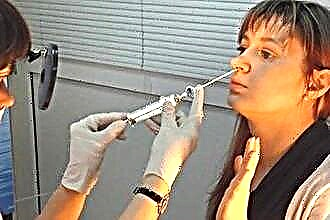
 With a visual examination of the nasal cavity, even with the help of an endoscope, it is impossible to accurately diagnose, to give any predictions for the further course of the disease, and even more so to prescribe treatment.
An interesting fact is that most often patients have an osteoma of the left frontal sinus. Today it is difficult to say what this is connected with, and why the tumor prefers to be localized on the left. But often it is found in children during a preventive medical examination, when a sharp decrease in vision in the left eye is detected, and doctors begin to find out its causes.
Treatment regimen
Based on the data obtained as a result of the examination, the doctor can make a final conclusion about the patient's condition and decide how much treatment is necessary in this case, and which one.
If the osteoma does not cause concern and was discovered by accident, then doctors recommend conducting a dynamic examination every few months.
The best option for dynamic examination is a computed tomogram, which allows you to fairly accurately determine the speed and direction of the growth of the osteoma. Sometimes it happens that she simply stops growing, although no treatment has been carried out. But more often it just increases so slowly that you can ignore it.
With a strong growth of the osteoma and infringement of adjacent tissues or organs, it is necessary to make a decision about its surgical removal (if at all possible, based on the localization of the tumor and the patient's state of health). Conservative treatment cannot slow down the growth of the osteoma; it can only be used to relieve the symptoms caused by the tumor.
 The operation is performed in a hospital under general anesthesia, since sometimes it is necessary to remove large areas of bone and tumor-damaged vessels and soft tissues. Therefore, before carrying out it, the patient must be carefully examined for the absence of medical contraindications to the use of anesthesia.
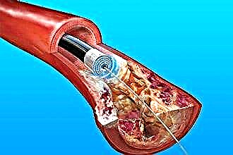
With a small size of the tumor and its suitable localization, a low-traumatic version of the operation is performed using laser endoscopy. Sometimes the surgeon has to open the frontal sinuses or perform craniotomy to gain access to the damaged area.
The active recovery period after surgery is up to 7-10 days. It is better for the patient to be in the hospital at this time in order to exclude the risk of postoperative complications or to quickly eliminate them.
To prevent the development of inflammatory processes, a course of antibiotic therapy is usually prescribed. The patient should follow a sparing diet, avoid drafts and sudden temperature changes. Reklama: stiklo konstrukcijos, pertvaros, turėklai, laiptai, durys ir terasos stogo stiklinimas gera kaina
Within 2-3 months after the operation, a number of restrictions are strictly observed: you cannot visit the pool, swim in open water, diving and active sports. It is very important to protect yourself from respiratory viruses and infections, since the disease during the recovery period can provoke various complications.
Prevention measures
It is difficult to advise any effective preventive measures that will prevent the development of osteomas. The best prevention of any neoplasm is a healthy lifestyle and strong immunity. Efforts should be directed towards achieving these goals.
 Particular attention should be paid to the diet: it should contain a sufficient amount of calcium. To do this, include in the daily menu: greens, sesame seeds, all types of dairy products, nuts. Vitamin D from tablets is absorbed by only 15%, but under the influence of the sun it is actively produced by the skin. Therefore, you need to try to spend at least half an hour a day on the street.
Frequent colds and chronic respiratory diseases weaken the immune system and lead to atrophic changes in the nasal cavity and sinuses. This also contributes to the development of all kinds of neoplasms, and not only benign ones. This means that you need to be treated with high quality and consult a doctor in a timely manner.
They talk about giving up bad habits everywhere and all the time. We have to repeat ourselves - smoking significantly increases the risk of cancer and the development of benign neoplasms in the respiratory organs.
By the way, the recovery period after surgery in smokers lasts longer and the risk of complications is higher. So decide for yourself.