The heart is rightfully defined as the most important organ of the human body: since ancient times, it was believed that the soul is located behind the sternum and leaves the body with the last blow. The organ is laid in the sixth week of intrauterine development. The importance of adequate functioning of all structures of the heart determines the length and quality of life of each person. Therefore, knowledge of the basic constituent anatomy and physiology of an organ is necessary for a clear understanding of possible problems and their consequences.
How does the human heart work?
The heart (Latin cor) is a muscular-cavity formation, which ensures an adequate supply of blood to all cells and tissues. The peculiarity of the organ is autonomy: individual innervation and regulation of the contractile function. However, the muscle, valve and structures of the conducting system are extremely sensitive to changes throughout the body.
Organ topography: the heart is located in the chest cavity in the complex of structures of the mediastinum (formation that is located between the two lungs), occupying the middle lower part. The organ "lies" on the diaphragm, enclosed in a pericardial sac - the pericardium. The lateral walls are adjacent to the roots of the lungs and the great vessels.
Schematic representation of the internal structure of the heart:
With a general clinical examination by percussion (tapping) on the anterior chest wall, relative and absolute cardiac dullness is determined. The predominant part of the organ is on the left side, the right border is along the outer edge of the sternum.
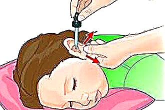
Listen to the activity of the heart, the functioning of the valves with a phonendoscope at the points of their projection.
Anatomy
The morphological structure of the heart is determined by experts in different ways. Anatomically, the organ is divided into the right and left halves, which are connected through the vessels of the large and small circle of blood circulation.
During intrauterine development, the heart goes through different stages of chamber formation. In the case of an incomplete process at birth, pathological shunts between the left and right sections persist, which cause hemodynamic disturbances.
 The chambers (cavities) of both halves are interconnected by means of holes, where the flow direction is regulated by the activity of the valve flap structures.
The organ wall is represented by three main sheaths:
- endocardium - lines the inner surface of the heart, forms tendon chords (threads) and valve apparatus;
- myocardium - the muscle layer that forms the wall of the organ, the interventricular septum and papillary muscles;
- epicardium - the outer connective tissue membrane, which is considered the inner layer of the pericardium. There is a small amount (up to 2 ml) of fluid between the layers of the pericardium, which ensures smooth sliding of the organ during different phases of the cardiac cycle.
Inflammatory pathologies of the pericardium or reactive changes in the background of other diseases (for example, pancreatitis or acute renal failure) lead to increased fluid synthesis, which prevents the expansion of the heart cavities and adequate blood flow.
Cameras
The diagram of the structure of the heart implies the division of the organ into halves, which are represented by four main and two additional chambers.
| Right part | Left departments |
|---|---|
| The atrium (atrium), which collects carbon dioxide-rich blood (venous) from the entire body | The atrium, where the four pulmonary veins flow, carrying arterial blood with a high concentration of oxygen |
| The ventricle, which is connected to the superior chamber through the atrioventricular opening. The outflow tract carries blood in a small circle for gas exchange | The ventricle is the largest chamber with a thick layer of muscle fibers, the contraction of which provides an adequate release of blood for delivery to the periphery |
| The ear is a small cavity connected to the atrium (smaller than on the left) | Ushko - additional chamber with an entrance to the atrium |
The clinical significance of the ears is the additional volume that fills the heart with increased loads. However, stagnation of blood in the chambers increases the risk of developing blood clots (clots) with possible spreading into the vessels of the brain or myocardium and subsequent stroke or heart attack.
Valve structures
 Regulation of blood flow in a certain direction is set by valve structures derived from the connective tissue inner membrane (endocardium). There are four main valves in the hemodynamic system of an organ:
- mitral (left atrioventricular) - represented by two valves that open into the cavity of the ventricles during atrial contraction;
- aortic (consists of three valves) - located at the exit of the left ventricle;
- tricuspid, which determines the movement of blood in the right sections;
- a pulmonary artery valve (tricuspid) that regulates the flow of fluid from the ventricle into the lesser circulation.
Closing and opening of the valve cusps is ensured by the contraction of the papillary muscles and the length of the tendon chords (too short or long fibers of the latter lead to a failure of the apparatus and reverse flow of blood).
Organ vascular system
Constant muscular work of the heart requires a large amount of energy, which is supplied through the coronary arteries with nutrients and oxygen. The coronary vessels of the organ are separated from the aorta directly at the base of the valve leaflets.
There are two main arteries supplying the myocardium:
- The right one extending from the aorta to the posterior surface of the heart provides trophism of the right atrium and ventricle.
- The left one, which bends around the atrium and lies in the anterior groove, provides blood supply to the main muscle mass of the heart (left sections, interventricular septum and anterior wall). Disruption of blood flow in this vessel most often causes pain and a tingling sensation behind the breastbone.
There are individual characteristics of the discharge of the arteries, therefore, with contrasting methods of research, different types of blood supply to the heart are distinguished.
The outflow of venous blood occurs through the vessels of the same name, which open with small holes into the cavity of the right atrium.
Histology: what does the heart look like under a microscope?
The structure of the heart is organized by three main membranes, the cellular structure of which is determined by the functions performed. The microscopic location of tissues in the section (histology) is presented in the table:
| Layer | Painting under a microscope |
|---|---|
| Endocardium (tissues of valves, tendon chords and papillary muscles, inner lining) |
|
| Myocardium | Muscle fibers built from mono- or binucleated cells. The contractile proteins have a transverse striation, as in skeletal muscles. The individual fibers are interconnected by means of insert discs. The latter contribute to the rapid spread of contraction throughout the mass of the heart muscle |
| Conductive system of the heart | There are three types of atypical cardiomyocytes (muscle) cells:
|
| Epicardium - the inner layer of the pericardium | A thin connective tissue sheath containing elastic and collagen fibers. |
The photo shows the histological structure of the heart (muscle layer):
Circles of blood circulation: where and from where does blood move through the vessels?
The main function of the heart is to provide adequate blood supply to all structures of the body. This task is realized with the help of coordinated work of the cardiovascular and respiratory systems.
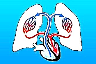
Schematic representation of blood circulation in the body:
In functional anatomy, two circles are distinguished along which the blood moves (large and small) and passes through the stages of providing the body with oxygen, nutrients and excretion of toxic metabolites (metabolic products).
Big circle
Arterial blood is transported along a large circle of circulation, starting from the cavity of the left ventricle. During the contraction of the latter, fluid enters the aorta - the largest vessel in the human body, individual branches of which deliver nutrients throughout the body:
- coronary vessels;
- subclavian artery, the branches of which feed the organs of the head, neck, structures of the upper limb;
- intercostal and bronchial, providing trophism of the mediastinal organs, lungs and structures of the chest wall;
- celiac trunk, renal and mesenteric arteries feed all organs of the digestive tract, urinary system, abdominal wall;
- bifurcation (bifurcation) of the aorta into the common iliac arteries provides trophism of the structures of the small pelvis and lower extremities.
Blood is transported through the vessels with a gradual narrowing of the diameter: from the arteries and arterioles to the capillaries. The cell wall of the latter has large pores through which oxygen and nutrients move to the tissues behind the concentration gradient.
Waste blood is taken in the end section of the capillary, then along the venules and to the main vena cava, which flow into the cavity of the right atrium:
- lower - from the structures of the abdominal cavity, small pelvis, soft tissues of the legs;
- upper - from the organs of the head and neck, part of the chest cavity.
Small circle
Venous blood entering the right heart is enriched with carbon dioxide, high concentrations of which have a depressing effect on the respiratory and vasomotor centers of the brain. Gas is excreted using the pulmonary circulation starting from the right ventricle:
 The pulmonary trunk, which divides into the right and left artery.
- Lobar and segmental arteries.
- Pulmonary capillaries, which are part of the air-blood barrier. The thin walls of the alveoli and blood vessels facilitate the movement of oxygen and carbon dioxide by a diffusion mechanism (concentration gradient).
- Venules that flow into the main veins (two from each lung) and carry blood to the left atrium.
 The pulmonary trunk, which divides into the right and left artery.
The name of the vessels is determined not by the composition of the blood, but by the direction in relation to the heart: fluid moves through the veins to the organ, along the artery from it.
Cardiac cycle
Adequate blood supply to the body is provided by a well-coordinated contraction of the muscle fibers of the heart wall, which determine the cycle of the organ.
There are two main phases:
- systole - contraction;
- diastole - relaxation.
Different speed of impulse conduction through atypical cardiomyocytes with a delay in the atrioventricular node ensures the coordinated work of the organ: during atrial systole, blood enters the ventricles. The latter are in the relaxation phase, which forms a sufficient volume for filling with liquid (in the left one up to 100 ml).
During the contraction of the ventricles, the valves of the aorta and pulmonary artery open, the valves of the atrioventricular joints are closed - the blood goes into circulation. On the peripheral vessels, the pulse is determined, and the heartbeat in the chest area.
At this time, the atria are in the diastole phase and are filled with blood from the hollow (right) and pulmonary veins (left).
There is a statement that the heart works half its life and half it rests, since the duration of systole and diastole is the same (0.4 seconds each).
Heart functions
The heart is rightfully considered the main organ of the human body, because the violation of its functions causes total disorders, and the cessation of activity leads to the death of the patient.
The main functions of the human heart:
 automatism - an independent synthesis of nerve impulses for the contraction of the myocardium;
- conductivity - atypical cells ensure the smooth functioning of different parts of the musculature of the organ;
- pumping function - pumping blood through the body with sufficient pressure to deliver it to the periphery;
- gas exchange is provided due to the work of a small circle according to the principle of an oxygen concentration gradient;
- endocrine role - natriuretic hormone is produced in the wall of the left atrium, which affects the functioning of the kidneys and the excretion of salts from the body.
 automatism - an independent synthesis of nerve impulses for the contraction of the myocardium;
Conclusions
The cardiovascular and respiratory systems are considered vital systems of the human body. The structure and functions of the heart directly determine the work of other organs due to an adequate blood supply to the brain, endocrine glands and kidneys.