The heart is the central organ of the cardiovascular system, providing blood circulation through rhythmic contractions and pushing blood out of the cavities into the great vessels. The correct direction of blood flow is carried out by the valve apparatus. The tricuspid valve, also called the atrioventricular opening (AV), is located between the right atrium and the ventricle. The main pathologies are congenital or acquired defects, complications after infectious diseases. If pathology is detected, treatment with a cardiologist is indicated.
How does the tricuspid valve work?
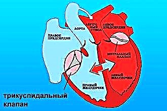
The human heart consists of four sections. The aortic valve and the pulmonary valve communicate with the atrial cavity with the arteries of the same name. The left and right atrioventricular foramen are located between the ventricle and the atrium of the heart on the corresponding side.
The tricuspid valve (right AV opening) is a window with three flexible plates - leaflets on a ring of fibrous tissue that communicates to the right heart. Normally, it has three valves: anterior, posterior and septal, due to which it is also called tricuspid. The presence of four or six leaves is possible.
The AV opening contains the papillary muscles and tendon chords extending from them, which are attached to the base of each leaflet and provide proper valve function, tension or relaxation during the heartbeat cycles. The right atrioventricular valve is depicted in the figure below:
Mechanism of work
During diastole (relaxation of the heart muscle), the tricuspid valve opens and allows venous blood to flow from the right atrium to the right ventricle. In systole (contraction of the heart), the flaps close tightly and do not wrap due to fixation with chords and muscles. From the ventricle, blood is released into the pulmonary trunk. Further, along the right and left main artery, it is sent to the lungs for gas exchange. At the same time, the muscles and functional apparatus of the valve prevent the return flow of blood into the cardiac cavity from large vessels.
This is due to:
- Keeping the valve leaflets open with the papillary muscles and chords during the filling phase.
- Tight closure to prevent regurgitation (reverse flow into the atrium) during the phase of expulsion of blood into the pulmonary trunk.
- Differences between the larger size of the valve cusps and the smaller diameter of the AV hole, which causes the valve to close tightly during the contraction phase when the volume of the ventricle changes.
- Anatomically, the valve is funnel-shaped, thereby providing passive blood flow when the pressure in the heart cavities changes.
Main functions
Throughout a person's life, the heart provides a closed path of blood flow, delivery of oxygenated blood to organs and tissues, venous outflow of carbon dioxide and decay products. The cardiovascular system consists of the circulatory system. The large one originates in the left ventricle and ends in the right atrium, the small one starts in the right ventricle and goes to the left atrium.
The tricuspid valve is actually a small circle element that performs the following functions:
- During heartbeats, it inhibits reverse regurgitation (blood flow from the lower ventricle to the atrium).
- Directly involved in blood circulation, provides delivery of venous blood to the vessels of the lungs.
- By means of which the process of gas exchange in the alveoli of the lung tissue and heat transfer are carried out.
What pathologies of the right atrioventricular valve most often occur
Dysfunction of the right atrioventricular foramen most often takes the form of stenosis or insufficiency. Pathological changes in the valvular apparatus of the heart significantly disrupt blood circulation, which is manifested by certain clinical symptoms.
Tricuspid valve stenosis
- Has a connection with some diseases of an infectious nature, infection with streptococcal, enterococcal or treponemal infections.
- It is more often detected in patients with rheumatism or syphilis.
- Represents a narrowing and a decrease in the diameter of the AV hole (stenosis), which significantly impedes the flow of blood through the valve.
- In 60%, it is combined with damage to other valves, mitral or aortic.
- Circulating in the bloodstream, the infection settles in all parts of the heart, affecting the elements of the valve apparatus.
- Due to the progressive inflammatory process, the tricuspid valve becomes sclerosed. The cusps, annulus fibrosus, muscle elements and chords grow together, reducing the lumen of the AV hole.
- Normally, the size of the valve is 3-4 centimeters, with stenosis, the diameter decreases from 3-1.5 cm.
- As a result of changes in hemodynamics, not all blood volume flows from the atrium into the ventricle, and therefore stagnation develops in the pulmonary circulation.
- During the examination, pathological reflux is characteristic - swelling of the cervical veins when pressed on the abdomen, where the liver is located.
- With auscultatory listening of the heart, diffuse pulsation and an increase in cardiac boundaries, a loud pathological noise in the diastole phase are revealed.
- It is manifested by portal hypertension (increased pressure in large hepatic vessels) with subsequent stagnation of blood in the spleen, intestinal vessels, and stomach.
- Typical symptoms of severe weakness, shortness of breath, edema, blueness of the hands and face, irregular heartbeat, increased pressure, hemoptysis, edema of the abdomen and fatty tissue, swelling of the veins around the navel.
- Leads to fatal heart failure and death if left untreated.
- Medical treatment is ineffective; surgical intervention is indicated to replace the affected valve and save the patient's life.
Insufficiency of the right atrioventricular valve
 Most often it occurs as a result of rheumatic infection, inflammation of the endocardium of the heart or rupture of elements of the valve apparatus, part of the chords or muscle fibers.
- It is extremely rare - it is a congenital defect.
- In addition to infection, changes in the right ventricle, its hypertrophy or dilatation can provoke dysfunction, which leads to a compensatory increase in the diameter of the fibrous ring of the valve and disrupts its closure.
- May occur as a result of expansion of the ventricle of the heart in inflammatory diseases, myocarditis or cardiomyopathy.
- Has a connection with opium addiction and its main complication - endocarditis (inflammation of the inner layer of the heart shell).
- It is characterized by incomplete collapse or prolapse (protrusion) of the valve leaflets, due to which the blood is constantly thrown back into the right atrium.
- When examining an ultrasound scan, the doctor sees a change in hemodynamics, the degree of blood flow and the size of the narrowing of the AV hole.
- With auscultation (listening to the valves with palpitations), a pathological clapping noise is determined.
- Since the atrium does not have great compensatory capabilities, signs of dilatation (expansion) immediately appear.
- As with stenosis, it is manifested by stagnation in the liver, an increase in venous pressure and a venous pulse (swelling of the neck veins when the heart contracts).
 Most often it occurs as a result of rheumatic infection, inflammation of the endocardium of the heart or rupture of elements of the valve apparatus, part of the chords or muscle fibers.
Consequences of disruption of the tricuspid valve
The prognosis for progressive stenosis or right atrioventricular valve failure is extremely poor. According to statistics, most patients develop complications of cardiac pathology within 5-10 years.The most dangerous of them: thromboembolism of the pulmonary vessels with a thrombus from the valve of the AV hole or fatal atrial fibrillation (arrhythmia). A complete blockade or a secondary infection in the presence of a defect can provoke a cardiac arrest.
The progressive pathology of the tricuspid valve is complicated by congestive heart failure with damage to the liver and blood vessels of internal organs. This increases the risk of developing gastrointestinal bleeding with portal hypertension from the veins of the esophagus.
Conclusions
The basis of prophylaxis for tricuspid valve pathology is timely examination and treatment. Drug therapy can only be effective in the early stages. With late detection of valvular dysfunction, surgery is indicated. As a rule, the life expectancy of a patient with tricuspid stenosis is no more than 20 years, with insufficiency - 25-30. Currently, a successful implantation, plastic, correction or valve replacement is performed by surgery, which allows preventing dangerous complications, death and prolonging the patient's life.