Heart defects are a large group of diseases characterized by a defect in any of its structures or large vessels.
What is heart defect
To understand what a heart defect is, you need to understand the basics of the anatomy of this organ and the principles of its work.
The human heart consists of 4 chambers - 2 atria and 2 ventricles. Blood moves from one chamber to another through the holes that have valves. From the left ventricle, blood is released into the systemic circulation (aorta), oxygenates all organs and tissues of our body, and returns to the left atrium through the vena cava. From there it goes to the right ventricle, then to the pulmonary artery in order to be enriched with oxygen in the lungs, and through the pulmonary veins it returns to the right atrium, then to the left ventricle. Then the cycle repeats.
To prevent mixing of arterial and venous blood inside the heart, the left and right sections are separated by atrial and interventricular partitions. To prevent backflow of blood (from the ventricles to the atria or from the aorta to the left ventricle), there are valves that open and close at a specific time.

All heart defects are divided into 2 types - congenital and acquired.
As the name implies, congenital defects appear in a person from his very birth, and acquired defects arise in the process of later life.
The incidence of congenital heart defects (CHD) is approximately 5-8 cases per 1000 children. Acquired heart disease (ACD) occurs in 100-150 people per 100,000 population.
For ease of understanding the differences between CHD and PPS, I note that at the first, an anomaly develops, deformation of the main vessels (aorta and pulmonary trunk) or a defect in the septa, and with acquired ones, the valves are affected. But such a division can be considered arbitrary, since valves can be damaged even with congenital defects.
All this leads to a violation of hemodynamics (normal blood flow) inside the heart, the predominance of blood filling in some chambers and the impoverishment of others. As a result, arterial blood mixes with venous blood, certain chambers overflow with blood, stretch, and their walls thicken. The filling of other parts of the heart, on the contrary, decreases in comparison with the norm.
Most people with heart disease get a disability class. They cannot live a full life, like all healthy people, they need to constantly observe some kind of restrictions. Even purely psychologically it is difficult.
On the question of the army - people with heart defects are categorized as “unfit” or “partially fit” for urgent military service.
Is it possible to die from pathology
Unfortunately, the fact of death from heart disease is quite possible. The statistics of deaths with congenital heart disease are rather sad. Without timely medical intervention, it occurs in 70-80% of cases.
People with PPP die in about 15-20% of cases. The main cause of death in heart disease is heart failure, i.e. deterioration of the main function of the "pump" - pumping blood.
Other causes of death include cardiac arrhythmias such as paroxysmal ventricular tachycardia, atrial fibrillation, and atrioventricular blockade. Due to atrial fibrillation, thromboembolism often occurs in the brain, leading to a stroke.
Possible causes of occurrence
Among the causes of acquired defects, the most common are:
- Rheumatism, or rather, chronic rheumatic heart disease, is an inflammation of its inner membrane (including the valve apparatus), which develops after a streptococcal infection (sore throat) suffered (mainly in childhood).
- Infective endocarditis is the gradual destruction of the heart valves due to the multiplication of bacteria on them. A drift of infection can occur when a carious tooth is removed, with poor antiseptic treatment of the skin during an injection, or using non-sterile syringes.
- Atherosclerosis and degenerative valve changes are common in the elderly.
Of the more rare causes, syphilis and systemic pathologies can be distinguished - rheumatoid arthritis, lupus erythematosus, scleroderma.
The specific etiological factor of congenital malformations is difficult to establish. It can be:
- hereditary mutations - Down syndrome, Patau;
- maternal diseases - diabetes mellitus, thrombophilia, systemic vasculitis;
- intrauterine viral infections - rubella, cytomegalovirus, chickenpox;
- bad habits - smoking, drinking alcohol during pregnancy;
- exposure to ionizing radiation;
- the use of drugs that adversely affect the development of the fetus - antineoplastic agents, sulfonamides, tetracyclines.
How to tell if you have a heart defect
To find out if a person has a heart defect, I am guided by the following data:
- symptoms and complaints that bother the patient;
- physical status - the patient's appearance;
- electrocardiography;
- echocardiography (ultrasound of the heart);
- chest x-ray.
Symptoms, signs and typical appearance of the patient
People with heart defects mainly suffer from signs of heart failure. They have difficulty breathing, especially at night, due to the horizontal position of the body and an increase in pressure in the vessels of the lungs. For the same reasons, they may be bothered by a paroxysmal cough.
Patients (especially with CHD) get tired very quickly, even after very little physical activity, they constantly want to sleep, they feel dizzy, and they may even faint.
Due to the enlarged liver, the patient feels heaviness or pulling / aching pains in the right hypochondrium. In the evening, the legs are very swollen. Often worried about aching pain in the left side of the chest, palpitations, discomfort in the chest. Lower respiratory tract infections persist in patients with certain CHD.
Often I notice the so-called "drumstick symptom" in people with heart defects. This is a thickening of the terminal phalanges of the fingers. This symptom indicates a long-term violation of blood circulation throughout the body.

Newborns and infants with CHD are stunted and underweight. Often their lips, nose, and fingertips become bluish (cyanosis).
There are specific symptoms of heart disease. For example, with coarctation of the aorta, due to its pronounced narrowing, the circulation of the head, arms and upper body remains at the proper level, while the lower parts of the body and legs are depleted in blood. This leads to the fact that the muscles of the upper shoulder girdle stand out against the background of the underdeveloped muscles of the lower extremities. And the false impression of "athletic build" is created.
Another example is mitral stenosis. In the later stages of this PPS, against the background of a general pallor of the face, a bright bluish-pink blush appears on the cheeks, while the lips and nose have a blue tint. This is called the facies mitralis, or mitral face.

I want to note that a person with PPS can feel quite healthy for a long time and not experience any pain or difficulty in breathing. This is due to the fact that the heart is trying to compensate for hemodynamic disturbances and at first it copes well with this. However, sooner or later these mechanisms are insufficient, and the disease begins to manifest itself clinically.
When I examine such patients, I manage to identify some pathological signs, for example, increased cardiac impulse of the left or right ventricle, tremor of the chest. During auscultation of patients with heart disease, I often hear murmurs at the projection points of the valves, septa, and carotid arteries; strengthening, weakening or splitting tones.
Instrumental diagnostics
The main instrumental research methods for the diagnosis of heart defects:
- Electrocardiography. On the ECG, I can see signs of hypertrophy of different parts of the heart by changing the height, width and shape of the teeth. Arrhythmias are often found (especially often - atrial fibrillation).
- Echocardiography is perhaps the main diagnostic method that allows you to reliably establish a heart defect. Echo-KG clearly recognizes the state of valves, partitions, wall thickness and volume of the chambers. In Doppler mode, you can see the direction of blood flow between the departments (regurgitation), measure the pressure in the pulmonary artery. If a defect is suspected, for a more detailed image, I prescribe a transesophageal echocardiogram (the transducer is placed in the esophagus just behind the heart).
- X-ray of the chest organs - the picture shows very clearly the bulging of the pulmonary artery trunk, an increase in the pulmonary pattern due to an increase in pressure in the vessels of the lungs, a change in the shape of the shadow of the heart, a usuration of the ribs (an uneven contour due to compression by intercostal arteries).
Types of vices and their differences
As already mentioned, all heart defects are divided into congenital and acquired. They differ from each other in pathophysiology, severity, human life expectancy.
There are many classifications of CHD, but most often clinicians use the Marder classification, dividing all CHD into defects with and without cyanosis (ie, "blue" and "white").
Table 1. Characteristics of the CHD
A type | Name | The distinguishing feature | The mechanism of hemodynamic disturbance |
CHD without cyanosis (pale type) | Ventricular and interatrial septal defects | "Heart hump" (protrusion of the anterior chest wall) due to a strong increase in the RV. Intense systolic murmur in the III-IV intercostal space to the left of the sternum | Discharge of blood from left to right. Overload of the LV, then the right heart. Rapid development of pulmonary hypertension due to reflex spasm of the pulmonary arteries |
Patent ductus arteriosus | Systole-diastolic murmur in the II-III intercostal space to the left of the sternum | Discharge of blood from the aorta into the pulmonary artery, increased blood flow in the small circle, overload of the left heart | |
Isolated pulmonary stenosis | Weakening of the II tone and rough systolic murmur over the PA valve | A sharp overload of the pancreas, depletion of pulmonary blood flow | |
Coarctation of the aorta | High blood pressure, "athletic physique" "Chilliness of the legs", weakening or pulsation in the arteries of the lower extremities, Usulation of ribs on X-ray, systolic murmur along the entire left edge of the sternum | Obstruction of blood flow through the narrowed area of the aorta, LV overload | |
CHD with cyanosis (blue type) | Transposition of great vessels | Severe general hypoxia (cyanosis, "drumsticks"), heart hump, loud I tone at the apex | Lack of oxygen in the organs through which the systemic circulation passes. |
The only ventricle of the heart | Signs of hypoxia, systolic murmur at the apex | Mixing of arterial and venous blood, increased pulmonary blood flow, rapid ventricular overload | |
Fallot's tetrad | A sharp weakening of the II tone over the pulmonary artery | Blood dump from right to left |
Acquired heart defects are divided into 2 types - stenosis, i.e. narrowing of the opening between the chambers, and failure, i.e. incomplete closing of the valve. All PPP boil down to the overflow of some heart chambers with blood and the impoverishment of others, with all the ensuing consequences.
The most common APS in adults is aortic stenosis (about 80%).
Combined defects can occur - when a person simultaneously has both insufficiency and stenosis of the valve. It is also quite common for me to see people who have multiple valves affected. This is called concomitant heart disease.
Table 2. Characteristics of PPP
A type | Name | The distinguishing feature | The mechanism of hemodynamic disturbance |
Mitral valve (MK) defects | Insufficiency of MK | Weakening of the I tone, systolic murmur at the apex | Reverse discharge of blood into the left atrium |
Mitral stenosis | Loud I tone, diastolic murmur at the apex. Facies mitralis. | Severe overload of the left atrium, its hypertrophy and expansion. Increased pressure in the pulmonary vessels due to reflex spasm | |
Aortic valve (AK) defects | Lack of AK | Increased pulse blood pressure, visible pulsation of the carotid arteries, protodiastolic murmur on AK | Distension of the left ventricle by the reverse flow of blood from the aorta |
Aortic stenosis | Pain resembling angina pectoris, constant fainting. Rough systolic murmur on AK extending to the carotid arteries | Deterioration of blood ejection into the aorta, left ventricular overload | |
Pulmonary artery (PA) valve defects | Insufficiency of aircraft | Weakening of the II tone on the LA valve, protodiastolic murmur in the II intercostal space to the left of the sternum | Reverse discharge of blood into the right ventricle |
LA stenosis | Amplification and splitting of the II tone. Pronounced pulsation of the right ventricle | Obstruction of the ejection of blood into the LA, overloading of the pancreas | |
Tricuspid valve (TC) defects | Lack of TC | Systolic murmur on the TC | Reverse discharge of blood into the right atrium |
TC stenosis | Amplification of I tone on TC | Right atrial overload, dilatation and hypertrophy |
How are heart defects treated?
Unfortunately, there is no medicine that can cure a person from heart disease. And all congenital heart diseases are treated only by surgery. An exception is patent ductus arteriosus, a congenital malformation that can be completely eliminated pharmacologically. But this is effective only in the first day of a person's life. To do this, I prescribe an intravenous injection of a non-steroidal anti-inflammatory drug (Ibuprofen, Indomethacin) for 3 days.
If there is cyanosis and signs of severe heart failure, surgery is performed immediately. Often, surgeons have to operate even on infants and one-year-olds. If the defect was found with instrumental methods of research, and the patient is not worried about anything, or there are minor symptoms, the operation can be postponed.
Traditionally, surgical interventions to eliminate CHD are carried out under general anesthesia, on an open heart, connected to a heart-lung machine. The defect is either sutured or closed with a pericardial or synthetic tissue patch. The open duct is ligated or transected.
Recently, in specialized cardiological centers with the appropriate equipment, it is possible to perform minimally invasive endovascular interventions. In such operations, under the control of ultrasound and X-ray, a catheter is inserted through the femoral vein, which reaches the right atrium. An occluder is inserted through the catheter, which is an interconnected disc of nickel-titanium wire. This occluder closes the defect.
The main contraindication to such operations is advanced pulmonary hypertension with severe vascular hardening. In these cases, the so-called palliative interventions are performed, which eliminate not the defect itself, but its consequences. Artificially created messages (anastomoses) between large vessels, so that the blood goes around the overloaded parts of the heart.
Now let's look at the treatment of acquired defects. With them, things are a little different.
If PPS developed against the background of rheumatism, then, according to the protocol, I will definitely use antibacterial therapy with penicillin antibiotics. This point is very important, since the presence of streptococcal bacteria in the body can cause the development of new heart defects.
Also, I always prescribe drug therapy that will help stabilize the patient's condition.
First of all, medications are used that slow down the progression of heart failure:
- ACE inhibitors - Perindopril, Ramipril;
- beta-blockers - Bisoprolol, Metoprolol;
- diuretics - Torasemide;
- aldosterone antagonists - Spironolactone, Eplerenone;
In case of heart rhythm disturbances, I use antiarrhythmic drugs - Sotalol, Amiodarone.
Anticoagulant therapy is also important, since part of the PPS, especially mitral stenosis, is often accompanied by atrial fibrillation, in which blood clots form in the left atrial cavity, leading to cardioembolic stroke. To prevent this, I prescribe warfarin or low molecular weight heparins.
When the patient is in serious condition, when the medications no longer help, I send patients for surgical treatment.
There are 2 main types of operations with PPP:
- valve replacement;
- reconstructive operations - valve plasty, commissurotomy, balloon valvotomy.
Valve prostheses are mechanical (artificial) and biological. Their key difference is as follows. When installing a biological valve, the patient should receive anticoagulant therapy for the first 3 months after surgery, and with implantation of a mechanical valve - for life. The choice of the type of valve is decided on an individual basis each time.
The only anticoagulant approved for long-term use in artificial heart valves is Warfarin.
Mechanical valves are more durable, but their cost is much higher than biological valves.
What determines the prognosis: how long do patients live?
I am often asked - "how long do they live with a heart defect?"
It depends on many factors, such as:
- kind of vice;
- its severity;
- the degree of heart failure;
- the presence of complications;
- timeliness of diagnosis and treatment;
- fulfillment of the doctor's recommendations (correct intake of drugs in compliance with all dosages, etc.);
- the quality of the operation performed.
Without surgery, patients with many CHD die in early childhood (up to 2-5 years). CHD, in which a person can live to adulthood without surgery, includes coarctation of the aorta, atrial septal defect.
The most favorable PPS in terms of prognosis is mitral, tricuspid regurgitation. Serious complications develop rarely and after a long time. With other ATS (mitral, aortic stenosis), patients die approximately 5-10 years after the first onset of symptoms.
Modern treatment options, both pharmacological and cardiac surgery, can extend the life of such people up to 60-70 years.
The consequences of pathology
A patient with heart disease, both congenital and acquired, has a high risk of developing acute heart failure (pulmonary edema, cardiogenic shock), which, without rapid medical intervention, leads to the death of a person.
Also, people with heart defects develop coronary artery disease much earlier, which means that they are several times more likely to develop myocardial infarction.
Almost any heart defect is accompanied by rhythm disturbances. The most dangerous of them are ventricular tachyarrhythmias and atrioventricular blockade.
With some defects, due to a pronounced overload of the pulmonary circulation and reflex vasoconstriction of the lungs, pulmonary hypertension occurs - a very serious condition that is difficult to respond to drug therapy, requiring surgical intervention.
Due to prolonged pronounced oxygen starvation (hypoxia) of the whole body, the immune system suffers, which is why patients with heart defects constantly suffer from infectious diseases, especially bronchitis and pneumonia.
With any heart defect, as well as the presence of prosthetic valves, the risk of infectious (bacterial) endocarditis, a dangerous disease that affects the heart valves, often ends in death, increases several times.
Case study: teenager with aortic coarctation
I will cite one interesting case from my practice. A mother came to me with her 15-year-old son, who has been bothered by headaches, chills and an incomprehensible weakness in the legs since early childhood. At the age of 7, the boy was in the hospital in the pediatrics department, where he was diagnosed with high blood pressure up to 150/90 mm Hg. Was diagnosed with "hypertension", prescribed drugs. The patient took medications irregularly. The patient's young age, as well as the absence of hypertension in the mother and father, made me doubt the diagnosis and suspect the “secondary nature” of high blood pressure.
During a general examination of the patient, in addition to increased blood pressure (155/90 mm Hg), I was able to identify a weakened pulsation in the arteries of the legs and a systolic murmur at the back at the level of the lower angle of the scapula. I ordered an echocardiogram, which showed a thickening of the left ventricle and an area of narrowing in the thoracic aorta. Another sign of coarctation of the aorta was clearly visible on the roentgenogram - usulation of the ribs (uneven contour). The patient underwent a surgical operation - plastic surgery of the narrowed area of the aorta. The boy's condition improved, blood pressure returned to normal, and the need for medications to correct blood pressure disappeared.
Expert Advice: Living With Heart Disease
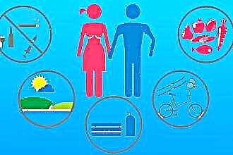
I want to give a few recommendations that will help to avoid most of the negative consequences and increase the effectiveness of treatment:
- sports - professional training will have to be stopped. Light physical activity is allowed;
- regular check-ups - it doesn't matter if you had a successful operation as a child or just recently diagnosed with mild mitral regurgitation. With a heart defect, it is necessary to visit a cardiologist at least once every six months or a year, to do an ECG and Echo-KG to check the state of heart functions, as well as to monitor the occurrence of possible complications;
- table salt - if you have been found to have signs of chronic heart failure and you have been prescribed medications for its treatment, for their greater effectiveness, you need to limit the use of table salt with food to 2-3 grams per day;
- Warfarin - This drug is often prescribed to prevent blood clots in patients with heart disease. In order for its reception to be effective and at the same time safe, you need to regularly do a blood test (coagulogram). The INR indicator in this study should be more than 2, but less than 3;
- visiting an otorhinolaryngologist - if you have been diagnosed with PPS of rheumatic origin, be sure to consult an ENT doctor, since the main cause of rheumatism is tonsillitis (tonsillitis). In the presence of chronic tonsillitis, treatment of the tonsils (washing, antibiotics) is necessary, and possibly their removal. This is necessary to prevent the recurrence of rheumatism and the appearance of a new heart defect.
- prevention of infective endocarditis - all people with heart defects and prosthetic valves have an increased risk of developing infective endocarditis. Therefore, to prevent it, they must take penicillin antibiotics (Amoxicillin, Ampicillin) once, about 30 minutes / 1 hour before medical procedures (tooth extraction, bronchoscopy, cystoscopy, etc.).