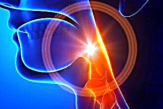
What is the pulmonary valve and where is it?
Pulmonary (PC) or pulmonary valve (PA) is an anatomical structure that separates the cavity of the right ventricle from the pulmonary trunk. The latter takes part in the formation of a small circle of blood circulation, delivering venous blood to the lungs through the system of small arteries and capillaries.
In relation to the external landmarks, the pulmonary valve is located in the area of attachment of the cartilage of the 3rd rib to the sternum on the left (the point of auscultation of the PC work).
Structural components:
- 3 semi-moon flaps (flaps): front, left, right;
- 3 sinuses ("sinuses");
- an annulus fibrosus, the main function of which is a supporting frame for the leaflets.
The flaps are shaped like pockets attached to the fibrous ring, the free edges of which are directed towards the lumen of the vessel. The lines of closure of the valves are called commissures. The thickenings in the central part of the valve are Morgagni's nodules.
The leaves are formed by three layers:
- ventricular (ventricular);
- middle (spongy);
- sinus (fibrous).
The thickest valve is at the point of attachment to the annulus fibrosus, narrowing in the central direction. Valvular structures are supplied with blood by separate arteries extending from the pulmonary trunk.
The histological structure of the annulus fibrosus and leaflets is similar to the structures of the endocardium - the inner connective tissue membrane of the heart. The upper layer is a thin epithelium that protects the surface of the PC from the formation of blood clots. The elastic and collagen fibers of the lower layer provide density and extensibility.
Sinuses (or sinuses) - the space between the valves and the vessel wall, the name of each corresponds to the forming flap. The section of the artery wall that forms the sinus has a developed middle layer of smooth muscle cells and connective tissue elements. The latter becomes thinner towards the base of the sinus due to a decrease in the number of myocytes.
The annulus fibrosus is a triangular strand of dense connective tissue, which consists of collagen (the main component), elastin and a small amount of cartilaginous elements. Ring structures continue in the middle layer of the valves.
In the process of embryogenesis, PC develops from the mesenchyme, being part of the "ventricular outflow tract".
Hemodynamics
The main function of the pulmonary valve of the heart is to provide unilateral blood flow in the pulmonary system and prevent return (regurgitation) from the pulmonary trunk to the right ventricle.
During the period of systole (the moment of contraction of the heart), the pressure in the pancreas exceeds the values in the arteries, so the valve is open, the flaps are pressed against the vessel wall. When diastole (relaxation) occurs, the blood rushes in the opposite direction, flows into the sinuses, as a result of which the valves close.
Normal performance
To study the functioning of the CLA at the primary stage, a general clinical examination of the patient is used. Typical signs of violations:
- dyspnea;
- pulsation of the cervical veins;
- pallor or cyanosis of the upper part of the body;
- on auscultation - additional noise at the point of valve projection.
The final diagnosis and the choice of adequate treatment tactics depend on the results of instrumental methods that objectively assess the state of this structure. The most common tests are electrocardiography (ECG) and echocardiography (echocardiography).
ECG
There are no specific electrocardiographic signs of PC defects. Changes in the ECG occur when symptoms of heart failure due to a valvular defect appear.
With regurgitation or stenosis of the pulmonary valve, hypertrophy of the right ventricle develops, which is established on the ECG in comparison with the state of the left sections.
Pronounced hypertrophy of the pancreas (weight more than the left):
- the ventricular QRS complex is represented by q-R or simply R in chest lead V1;
- in V6 the QRS complex is deformed into r-S, R-S (occasionally - R-s);
- the degree of hypertrophy reflects the height of the R wave in V1 and depth S in V6 - the greater the difference between them, the more significant the increase in the myocardium;
- ST segment in V1 is under the isoline, the T wave is negative;
- in V6 ST is located above the isoline, T is positive.
Moderate hypertrophy (the mass of the pancreas is less than the left, but the impulse conduction is slowed down):
- QRS complex in V1 observed as rsR or rSR;
- V6 deformation in qRS.
Moderate hypertrophy is represented by deformation of the QRS complex:
- in V1 - rS, RS, Rs;
- in V6 - qR;
- amplitude of the R wave in V1 increased;
- S waves in V1 and R in V6 - reduced.
A shift of the electrical axis of the heart to the right is noted at each stage of hypertrophy.
ECHO
Cardiac ultrasound is considered the gold standard for diagnosing PC defects. The method allows you to study the anatomical features and functional state of the valves. Doppler echocardiography is used to visualize internal hemodynamics.
The pulmonary valve on the ECHO is depicted in the form of a three-lamellar formation, the elements of which during pancreatic systole are bent into the lumen of the vessel.
PC normative indicators:
- structure - normoechoic, homogeneous;
- peak blood flow velocity - 0.5-1.0 m / s;
- the average pressure gradient is up to 9 mm Hg. Art .;
- the diameter of the lumen is 11-22 mm.
During ultrasound, the structure of the CLA, the presence of pathological formations are studied. The main changes for different defects are presented in the table.
| CLA stenosis | Insufficiency (regurgitation) of CLA |
|---|---|
|
|
Conclusions
The pulmonary valve plays a significant role in maintaining normal hemodynamics of the pulmonary circulation and the activity of the right heart. In the case of acquired, genetically determined defects, deficiency or stenosis of the CLA occurs, which contributes to the development of right ventricular hypertrophy and dilated cardiomyopathy. The gold standard for the diagnosis of pathologies is echocardiography.


