The disease, which is better known as acoustic neuroma, has alternative names: vestibular (or acoustic) schwannoma and acoustic neuroma. Symptoms in 95% of cases begin to manifest themselves in the form of progressive hearing impairment, which in 60% is accompanied by noise or ringing in the ears. The difficulty of detecting the symptoms of acoustic neuroma and treating the disease is that the slow growth of the tumor causes the presence of a long asymptomatic period, as well as the gradual appearance of clinical manifestations.
Discovery and Study History
 In 1777, Sandyfort performed an autopsy, which resulted in a description of a dense benign tumor of the auditory nerve, which turned out to be spliced at the exit site of the vestibular cochlear and facial nerves with the brain stem and spread into the auditory canal of the temporal bone. The researcher concluded that this formation was the cause of the patient's deafness during life.
In 1830, Charles Bell was the first to diagnose a patient during his lifetime, which was confirmed after the patient's death. Bell focused on frequent headaches, deafness, loss of gustatory sensitivity, trigeminal neuralgia, and some other signs and complaints from a patient who died a year after the examination.
The first surgical operation to remove the encapsulated formation of the cerebellopontine angle was performed in 1894 by Charles Ballance. Despite the fact that during the operation, the trigeminal and facial nerve structures were damaged and because of the complication, enucleation of the eye was required, the medical intervention could be called successful, since after it the patient lived for more than 12 years.
Harvey Cushing, the founder of neurosurgery in the United States, made a difficult path to reduce postoperative mortality. After the first operation in 1906, which ended in the death of the patient, the neurosurgeon first abandoned the complete removal of the neuroma, which made it possible to reduce the mortality rate by up to 40%. And later, with the improvement of the technique, the postoperative mortality rate was reduced to 7.7%. However, out of 176 neuromas removed by Cushing, only 13 cases represented total removal.
Since 1917, Cushing's follower Walter Dandy has perfected the technique by using a suboccipital approach to the posterior fossa, which became possible through penetration through part of the occipital bone. As a result, the mortality rate dropped to 2.4%. However, to this day, surgical intervention for acoustic schwannoma is associated with serious risks associated with the health of patients.
Therefore, the introduction of radiosurgery by Lars Lexell made it possible to dramatically improve the quality of treatment for neurinomas up to 3 cm in size.
Morphology, causes and mechanisms of the onset of acoustic disease
The neoplasm is described as a rounded (or irregularly shaped) tuberous dense node that has a connecting capsule on the outside, and diffuse or local cystic cavities with a brownish fluid inside. Depending on the blood supply, the color of the neoplasm may be:
- pale pink with reddish patches (most often),
- cyanotic (with venous stasis),
- brown-brown (with hemorrhages).
A benign formation consists of cells that form a structure like palisades, between the elements of which there are sections that consist of fibers. In the process of proliferation, tissues become denser, deposits of pigment consisting of iron oxide (hemosiderin) appear.
VIII pair of FMN includes the vestibular part (carrying information to the cerebral centers from the vestibular receptors) and the auditory part. In most cases, a neuroma occurs in the vestibular part, squeezing adjacent sections as it grows. Since a number of others pass next to the vestibular cochlear nerve (ternary, abducers, vagus, glossopharyngeal, facial), their compression is also reflected by characteristic signs.
The causes of this benign formation are not fully determined.
There is no direct connection between unilateral neurinomas and etiofactors. The bilateral form is stably fixed in patients with genetically determined type II neurofibromatosis. There is a 50% risk of disease in offspring if the parents have an abnormal gene.
There are three stages of vestibular schwannoma growth:
- The first is characterized by the size of education up to 2-2.5 cm, entailing hearing loss and vestibular disorders.
- For the second, it grows to a size of 3-3.5 cm ("walnut") with pressure on the brain stem. This contributes to the onset of nystagmus and imbalance.
- The third stage occurs when the formation grows to the size of a chicken egg with symptoms of compression of cerebral structures, impaired swallowing and salivation, and visual function. At this stage, irreversible changes occur in the brain tissues, and, due to the fact that the schwannoma becomes inoperable, death occurs.
Symptoms
 With the typical development of the disease, hearing impairment (in 95% of patients) and dizziness (less often) are recorded as the first signs. Most often (in 60% of cases) acoustic effects in the form of noise, ringing or hum on the side of the neoplasm become the only symptom of the initial stages of the disease. It happens that hearing loss occurs noticeably and sharply. However, hearing impairment can manifest itself almost imperceptibly to the patient, and the noises subside with the appearance of other symptoms.
At the initial stage, vestibular disorders are also recorded in two out of three cases. They manifest themselves:
- dizziness, which with this disease manifests itself gradually with an increase in intensity,
- instability when turning the head and body,
- nystagmus, which is more pronounced when looking towards the location of the neoplasm.
Most often, parts of the auditory and vestibular nerve structures are affected at the same time. However, in some cases, only one of the parts may be affected.
Sometimes vestibular crises are recorded, characterized by nausea and vomiting against a background of dizziness.
With an increase in education and damage to nearby structures, signs of this lesion are added to the symptoms. However, the size of the neoplasm does not always correspond to the severity of the symptoms. In addition to the size, the degree depends on the direction of growth and localization of the schwannoma. So, with a large size, a schwannoma may show dimmer symptoms than a small one, and vice versa.
Compression of the trigeminal nerve structures causes aching pains on the face and sensations of numbness and tingling from the side of the neoplasm. Such pains can subside or intensify, and then become permanent. Sometimes this pain is confused with a toothache or mistaken for trigeminal neuralgia. It happens that pain occurs in the back of the head from the side of the formation.
In parallel with these processes (sometimes a little later), symptoms associated with peripheral lesions of the facial and abducens nerves appear:
- facial asymmetry due to paresis of facial muscles,
- loss of taste in 2/3 of the front of the tongue,
- violation of salivation,
- convergent strabismus,
- double vision with displacement (diplopia).
If a benign formation grows in the internal auditory canal, then the symptoms of squeezing can appear already in the first stages of the disease.
A further increase in education leads to impaired laryngeal functions, disorders of swallowing and pharyngeal reflex, loss of sensitivity in the rest of the tongue.
The late symptoms of the disease include an increase in intracranial pressure, which results in a violation of visual function, the total nature of a headache with a concentration in the occipital and frontal regions. Systematic vomiting occurs.
Diagnostics
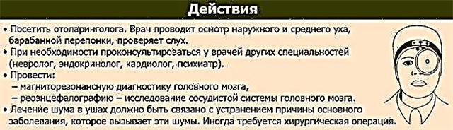
At the initial stage of the disease, it should be separated from similar manifestations of labyrinthitis, Meniere's disease, cochlear neuritis, otosclerosis. Diagnosis of the disease is carried out by an otoneurologist or at a joint appointment with a neurologist and an otolaryngologist. As needed, a vestibulologist, an ophthalmologist and sometimes a dentist are involved in the examination.
The examination begins with a check of the nervous system and hearing, and if a schwannoma is suspected, a number of diagnostic procedures are prescribed, which make it possible to confirm the suspicion with a high degree of probability:
- A pure tone audiogram is required for the initial determination of a treatment strategy and data collection for subsequent comparison.
- Electronystagmography. Effective use is possible only with a pronounced form of the disease, since while the neuroma is localized in the lower part of the vestibular apparatus, the method does not allow it to be detected.
- CT scan of the brain. When the size of the neoplasm is up to 1 cm, it is difficult to detect with the help of a CT picture. And with a size of up to 2 cm, only 40% of tumors are detected, unless contrast agents are specially introduced to facilitate diagnosis. When staining markers are introduced into the blood, they are absorbed by the neoplasm, and the X-ray density increases several times. The detected formations are more often rounded and have smooth outlines.

- MRI reveals a smooth contour of the neoplasm with deformation of the cerebellum, brainstem and a strip of the signal "CSF gap" along the periphery of the tumor.
- Radiography according to Stenvers with a snapshot of the temporal bone. An increase in the width of the internal auditory canal becomes a sign of the development of the disease.

Treatment by medical and folk methods
The outcome of treatment depends on the timely detection and size of the tumor. When a neuroma is diagnosed in the first two stages, the prognosis is favorable. Radiosurgical removal of the tumor stops the resumption of growth in 95% of cases. At the same time, the patient's working capacity is restored, he returns to his previous way of life. Open surgery has a less favorable prognosis associated with the risk of damage to various nerve structures and / or hearing loss. In the third stage of the disease, the prognosis is unfavorable. Vital cerebral structures are at risk of compression. When treating neuroma of the auditory nerve with folk remedies, predictions are not made, due to the lack of evidence of the effectiveness of folk methods.
Medical Approach
Since each of the treatment methods has both advantages and risks, they speak not about the only possible approach, but about the possible tactics of medical control and intervention.
- Expectant management involves monitoring the state of hearing with the help of audiometry and changes in symptoms. Tumor growth is monitored using MRI and CT: during the first two years, once every six months, then once a year. In case of unstable education behavior, the examination schedule is changed. As a rule, the same tactic is used if neuromas are detected by chance on MRI, when the development of the pathology passes without clinical symptoms. It is also used when monitoring elderly people with long-term hearing impairment.
- Tactics aimed at alleviating the patient's condition. For this, anti-inflammatory, analgesic and diuretic drugs are used.
- The use of radiation therapy allows the treatment of acoustic neuroma without surgery. The tumor cannot be removed with the help of radiation, but it can stop its growth and thereby avoid surgery.
- Stereotactic radiosurgery (SRS) involves the removal of neuromas up to 3 cm in size.It is also indicated for the elderly after subtotal resection (with prolonged growth) and in cases where the risk of open surgery due to somatic pathology is increased.
- Open surgery to remove a tumor is prescribed when it grows to a large size or has a noticeable growth dynamics, as well as in cases where radiosurgery has not helped.


The decision on surgical removal is made on the basis of a combination of accounting factors, including: the size of the tumor, the patient's age, the quality of hearing, the degree of qualification of the surgeon. Depending on the pathways of access to the neoplasm, the following types are distinguished:
- Suboccipital. The operation is performed with a high chance of preserving hearing.
- Translabyrinthine. There are several options, all of which carry a high risk of hearing damage.
- The infratemporal pathway is applicable when removing small neuromas through the middle cranial fossa (MF).
Traditional methods
In cases where it is not possible to carry out an operation in order to inhibit tumor growth and reduce its size, herbal therapy is used, which, nevertheless, cannot replace medical methods:
- Infusion of white mistletoe. The shoots of the plant are crushed, 2 teaspoons (with a slide) are poured with 2 cups of boiling water and infused overnight in a thermos. It is drunk three times a day, 2 tablespoons in small sips before meals. The duration of the course is 23 days. After a week, you can take the next course. (The maximum number of courses with a week break is four).
- Alcohol tincture of Japanese Sophora. For a liter of alcohol, 100 g of a ground plant is taken. Infusion occurs for 40 days with daily shaking (stirring). Next, the alcohol is filtered, and the cake is wrung out. It is taken 10 g three times a day before meals for 40 days. The break between courses is half a month.
- Horse chestnut vodka. The proportions and procedures are the same as in the previous case, but the composition is infused only for 10 days and in a dark place. It is drunk with a small amount of water, 10 drops three times a day. The 14-day course can be repeated for three months with a week break.
- Infusion of the Siberian prince. A teaspoon of a ground dried plant is infused for an hour in boiling water (2 glasses) and, after straining, is taken three times a day, a tablespoon for two months.
- Mordovan ordinary tea leaves. For brewing, crushed seeds are needed at the rate of a teaspoon per 300 ml of water. The composition is boiled for 15 minutes over low heat and infused for 2 hours before straining. It is consumed four times a day for 2 tablespoons.
- Common comfrey lotions. Powdered plant roots in a 1: 5 ratio are mixed with pork fat. Simmer this mixture for 5 hours in an oven preheated to 70C. Without letting the mixture cool down, it must be filtered and laid out in glass jars, tightly corked, and then twice a day a layer of ointment applied to parchment paper should be used as a local lotion (30 minutes / procedure). The monthly course of treatment is alternated with a two-week pause.